Papilio5D
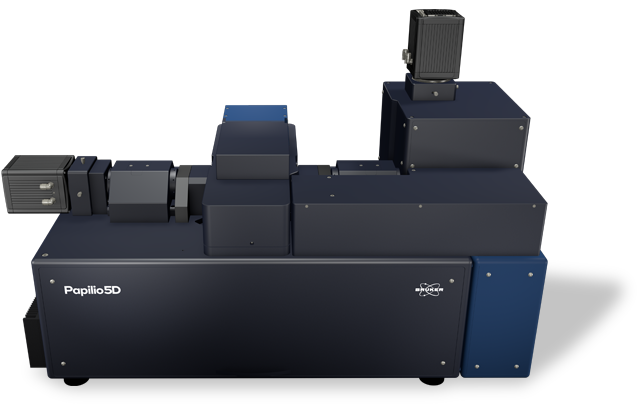
Papilio5D
The Bruker Papilio5D light-sheet microscope delivers high-sensitivity imaging for organoids and 3D cell cultures with a high degree of flexibility, enabling researchers to adapt the system to specific sample types, study conditions, and experimental designs.
The system’s horizontal lens geometry with dual-view detection and a choice of Gaussian or Bessel beam illumination, combined with support for simultaneous multi-sample and multi-condition imaging, make Papilio5D a flexible solution for achieving deep and high-resolution imaging while maintaining sample integrity — even on challenging samples and in complex, multi-sample, and multi-condition experiments.
To learn more, continue reading, contact us, or download the brochure.
Fast and Photo-Efficient for the Highest Quality Images
Papilio5D is photo-efficient for the most delicate samples, using a horizontal geometry with two views/dual-sided detection and a perpendicular light-sheet. This setup is capable of capturing twice as many photons as single-objective detection setups and enables a more comprehensive 3D view of samples.
Fast dual-view acquisition and fusion deliver high-resolution images. Water-dipping high-NA lenses ensure the light sheet is ultra-thin. Area- and line-mode detection also allow data acquisition specialized for different sample requirements.
Together, these features deliver:
- A high signal-to-noise ratio
- An impressive z resolution
- Optimized depth penetration
Papilio5D can also reach Nyquist sampling, which improves subsequent image processing and enables state-of-the-art deconvolution. Destriping further enhances image quality.

Customizable for Complex Experimental Design
Papilio5D was designed with maximum flexibility in mind. This includes multi-beam and multi-acquisition modes, as well as multi-sample mounting and multi-condition comparison.
Modular illumination beam options
Papilio5D users get to choose the best light beam for their experiment. The choice of Gaussian or Bessel beam illumination, both with a beam width below one micrometer, ensures outstanding image quality and great depth-penetration for a wide variety of objects, including dense organoids or zebrafish larvae and fish.
- Gaussian beams are ideal for small, light-sensitive samples, such as early mouse embryology studies.
- Bessel beams provide multi-direction illumination from a 360° angular range for robust imaging of even optically dense objects.
Multi-acquisition modes
These beams are complemented by two modes of acquisition: area or line mode.
- In area mode, the camera captures the entire field of view at once, making it fast and efficient.
- In line mode, the camera uses a confocal slit-like rolling shutter that synchronizes with the scanned light-sheet. This effectively rejects out-of-focus and scattered light, improving contrast in thick or scattering samples (e.g., organoids).

Multi-sample and multi-condition configurations
The Papilio5D has a generous sample holder that can hold up to three dishes with two separate chambers each, enabling a total of six experimental conditions to be tested at once. Sample health and integrity are further maintained with environmental control that supports long-term imaging (hours to days). Papilio5D provides control over temperature (20 to 37°C), gas concentration (CO₂ and O₂), and humidity.

Configuration options
| Illumination Modes | Gaussian Beam | Bessel Beam |
|---|---|---|
| Beam Waist Thickness [µm] | 1 bis 6 | 1 (main lobe) |
| Beam Length [µm] | 20 | 350 |
| Pivot Scan | ✓ | ✓ |
| Confocal Line Mode | ✓ | ✓ |
| Acquisition Options | - Gaussian beam - Gaussian with line mode - Gaussian with line mode and pivot scan | - Bessel beam - Bessel beam with line mode - Bessel beam with line mode and pivot scan |
| Illumination Optics | 20x 0.6NA | |
| Detection Optics | Dual 25x 1.1NA / 16x 0.8NA | |
| Multi-Sample/Multi-Condition Experiments | 6 seperate compartments | |
| Magnification Changer | 4 seperate steps | |
| Data processing | Content based dual view registration & fusion | |
| Photomanipulation | CW or pulsed, VIS to NIR | |
Optimized for Different Applications
Papilio5D supports a wide range of biological imaging applications, from deep imaging of complex 3D organoids to long-term in vivo studies. Its dual-view detection and multi-sample mounting enable comparative experiments, such as tracking regenerative processes in zebrafish under different treatment conditions.
TO DISCUSS YOUR SPECIFIC APPLICATION, CONTACT US.
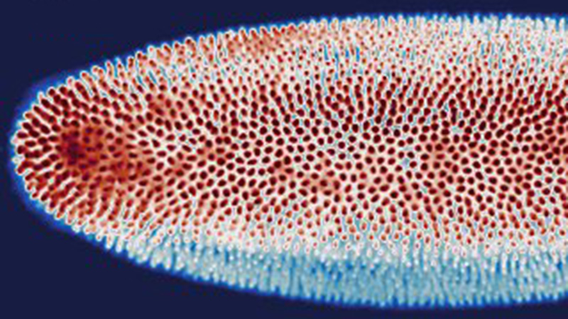
In Vivo Time-Lapse After Photomanipulation in Zebrafish
Time-lapse imaging of zebrafish distal tubules after localized photodamaging can provide novel insights into understanding wound healing processes over time. The design of Papilio5D sample mounting within separate chambers enables imaging under different treatment conditions to be studied at the same time. This allows comparative insights into how pharmaceutical components might impact wound healing.
In this example, regeneration of the distal tubule is studied in the embryonic zebrafish kidney using the cdh17:egfp transgenic line after photodamaging with a photomanipulation module.
Select Papilio5D Technical Specifications
| Dual Sided Detection with 2x Objectives | Tube Lens | Magnification | Pixel Size [nm] Orca Flash | FOV [µm] | Pixel Size [nm] Orca Fire | FOV [µm] Orca Fire |
|---|---|---|---|---|---|---|
| Detection 2x Nikon CFI APO LWD 25x 1.10 NA | 100 | 12.5x | 520 | 1065 | 370 | 876 |
| 200 | 25x | 260 | 532 | 184 | 436 | |
| 300 | 37.5x | 173 | 353 | 123 | 290 | |
| 400 | 50x | 130 | 266 | 92* (Nyquist sampling) | 218 | |
| Detection 2x Nikon CFI APO LWD 16x 0.8 NA | 100 | 8x | 813 | 1664 | 575 | 1362 |
| 200 | 16x | 406 | 832 | 288 | 681 | |
| 300 | 24x | 271 | 555 | 192 | 454 | |
| 400 | 32x | 203 | 416 | 144 | 340 | |
| DOWNLOAD THE BROCHURE TO LEARN MORE | ||||||
System Comparison
* COMPATIBLE MODULE
| The perfect allrounder - multi-view imaging for diverse samples (live and cleared option) | Multi-sample and dual view flexibility for highly sensitive imaging, ideal for organoids and 3D cell cultures | In vivo multi-sample and multi-condition imaging, ideal for drug treatment comparison | Perfection for cleared samples prepared by any type of clearing protocol | High-end imaging with various beam patterns from single cells to 3D cell cultures | |
|---|---|---|---|---|---|
| Geometry | Horizontal with multiview | Horizontal with dual view | Inverted with dual illumination | Inverted dual illumination with moving optics | Inverted geometry at 360° |
| Beam Type | Gaussian | Gaussian and Bessel | Gaussian | Gaussian | Advanced Illumination Module (AIM) |
| Application Examples | - Long-term imaging of Drosophila development - Photomanipulation studies in zebrafish embryos - Cleared and stained samples up to mouse brain size | - High-resolution full 3D imaging of multiple organoids over time - Long-term imaging of highly light-sensitive samples - Comparing the pharmacological impact in zebrafish wound healing after PM | - Time-lapse imaging of multiple pancreatic spheres - Studying pharmacological impact on zebrafish development - Comparing genetically altered organoids | - Pharmacokinetics in mouse brain - Multi-stained rat brain - Whole mouse imaging | - Fast imaging of photo-sensitive 2D cell culture - Lattice imaging for enhanced resolution in 3D cell cultures - FCS and FLIM in cell culture |
| User Level | Beginner friendly | Beginner friendly | Beginner friendly | Intermediate | Advanced |
| # Lenses | 4 lenses (2 IO, 2 DO) | 3 lenses (1 IO, 2 DO) | 3 lenses (2 IO, 1 DO) | 3 lenses (2 IO, 1 DO) | 2 lenses (1 IO, 1 DO) |
| Multi-View | ✓ | ✓ | |||
| Live/Fixed Samples | ✓ | ✓ | ✓ | ✓ | |
| Cleared Samples | ✓ | ✓ | |||
| Expanded Samples | ✓ | ✓ | ✓ | ✓ | ✓ |
| Best Embedding | Capillary/FEP tube w/ agarose 3D stage | Custom-solution and FEP foil | TruLive3D dishes and FEP foil | Quartz-crystal cuvette | FEP foil; glass slides |
| Photomanipulation* | ✓ | ✓ | ✓ | ✓ | |
| Environmental Control* | ✓ | ✓ | ✓ | ✓ | |
| Destriping/Uniform Illumination* | ✓ | ✓ | ✓ | ✓ | ✓ (w/ Advanced Illumination Module) |
| Benchtop Design | ✓ | ✓ | ✓ | ✓ | ✓ |
All-in-One LuxBundle Software
Intuitive design
Bruker's LuxBundle software saves time and enhances productivity by providing:
- All-in-one, easy-to-use interface for acquisition, viewing, and post-processing
- Fully scriptable microscope control and post-processing via open interface (e.g., Python or any other language), ready for custom "smart" microscopy
- High reproducibility of experiments: all parameters are saved in the metadata and configurations can be saved for future experiments
- Data formats (.tiff, .hdf5, .ims) compatible with common image processing software: Imaris, Aivia, BigDataViewer, Arivis, Fiji, Python, Matlab, Napari
Impressive Image Post Processor
Our dual-view light-sheet microscopes record a sample from different angles/views and generate images composed of multiple tiles. The LuxBundle software ensures high-quality, 360° crisp images of the sample that compensate for absorption and scattering. Features include:
- Multi-color alignment
- Tile stitching of hundreds of tiles for large samples
- Multi-view image fusion and deconvolution
3D Data Viewer
LuxBundle's integrated 3D data viewer allows researchers to inspect the entire dataset directly after acquisition. This gives users control over their data with key capabilities, including:
- The ability to turn tile stitching on or off
- Both raw and post-processed images
- Fast viewing of multi-terabyte data sets
- Flexible options to draw and annotate regions and landmarks