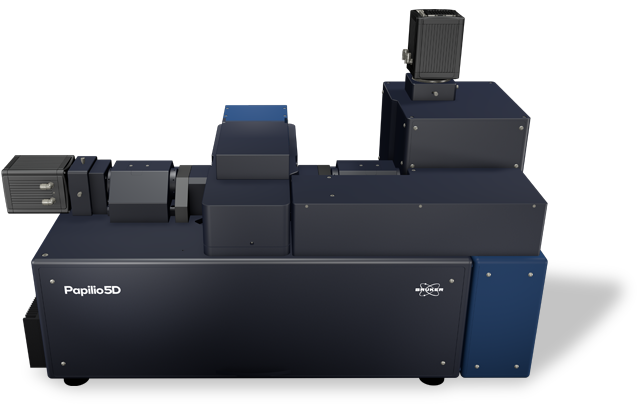
Luxendo Light-Sheet Microscopes

Papilio5D

Multi-sample and dual-view light-sheet microscope ideal for organoids and 3D cell cultures

Papilio5D

The Bruker Papilio5D light-sheet microscope delivers high-sensitivity imaging for organoids and 3D cell cultures with a high degree of flexibility, enabling researchers to adapt the system to specific sample types, study conditions, and experimental designs.

The system’s horizontal lens geometry with dual-view detection and a choice of Gaussian or Bessel beam illumination, combined with support for simultaneous multi-sample and multi-condition imaging, make Papilio5D a flexible solution for achieving deep and high-resolution imaging while maintaining sample integrity — even on challenging samples and in complex, multi-sample, and multi-condition experiments.

To learn more, continue reading, contact us, or see FAQs about this product.

Unprecedented
photon efficiency
With dual-sided detection and modular illumination beams.
Fast
dual-view acquisition and fusion
For highest-quality, high-resolution images.
Delicate & simultaneous
light-sheet imaging
For diverse samples under different experimental conditions.
Features

Fast and Photo-Efficient for the Highest Quality Images

Papilio5D is photo-efficient for the most delicate samples, using a horizontal geometry with two views/dual-sided detection and a perpendicular light-sheet. This setup is capable of capturing twice as many photons as single-objective detection setups and enables a more comprehensive 3D view of samples.

Fast dual-view acquisition and fusion deliver high-resolution images. Water-dipping high-NA lenses ensure the light sheet is ultra-thin. Area- and line-mode detection also allow data acquisition specialized for different sample requirements.

Together, these features deliver:

  • A high signal-to-noise ratio
  • An impressive z resolution
  • Optimized depth penetration

Papilio5D can also reach Nyquist sampling, which improves subsequent image processing and enables state-of-the-art deconvolution. Destriping further enhances image quality.

Papilio5D uses horizontal lenses, dual-view detection, and Bessel or Gaussian illumination. This setup enables optimized depth penetration and provides a large and evenly illuminated field of view with four different optical magnifications.

Customizable for Complex Experimental Design

Papilio5D was designed with maximum flexibility in mind. This includes multi-beam and multi-acquisition modes, as well as multi-sample mounting and multi-condition comparison. 

Modular illumination beam options

Papilio5D users get to choose the best light beam for their experiment. The choice of Gaussian or Bessel beam illumination, both with a beam width below one micrometer, ensures outstanding image quality and great depth-penetration for a wide variety of objects, including dense organoids or zebrafish larvae and fish.

  • Gaussian beams are ideal for small, light-sensitive samples, such as early mouse embryology studies.
  • Bessel beams provide multi-direction illumination from a 360° angular range for robust imaging of even optically dense objects.

Multi-acquisition modes

These beams are complemented by two modes of acquisition: area or line mode.

  • In area mode, the camera captures the entire field of view at once, making it fast and efficient.
  • In line mode, the camera uses a confocal slit-like rolling shutter that synchronizes with the scanned light-sheet. This effectively rejects out-of-focus and scattered light, improving contrast in thick or scattering samples (e.g., organoids). 
In this example of organoid imaging, all four Gaussian beam, Bessel beam, area and line modes are compared, illustrating different penetration depths.

Multi-sample and multi-condition configurations

The Papilio5D has a generous sample holder that can hold up to three dishes with two separate chambers each, enabling a total of six experimental conditions to be tested at once. Sample health and integrity are further maintained with environmental control that supports long-term imaging (hours to days). Papilio5D provides control over temperature (20 to 34°C), gas concentration (CO₂ and O₂), and humidity. 

Applications

Optimized for Different Applications

Papilio5D supports a wide range of biological imaging applications, from deep imaging of complex 3D organoids to long-term in vivo studies. Its dual-view detection and multi-sample mounting enable comparative experiments, such as tracking regenerative processes in zebrafish under different treatment conditions.

TO DISCUSS YOUR SPECIFIC APPLICATION, CONTACT US.

Case Study 1

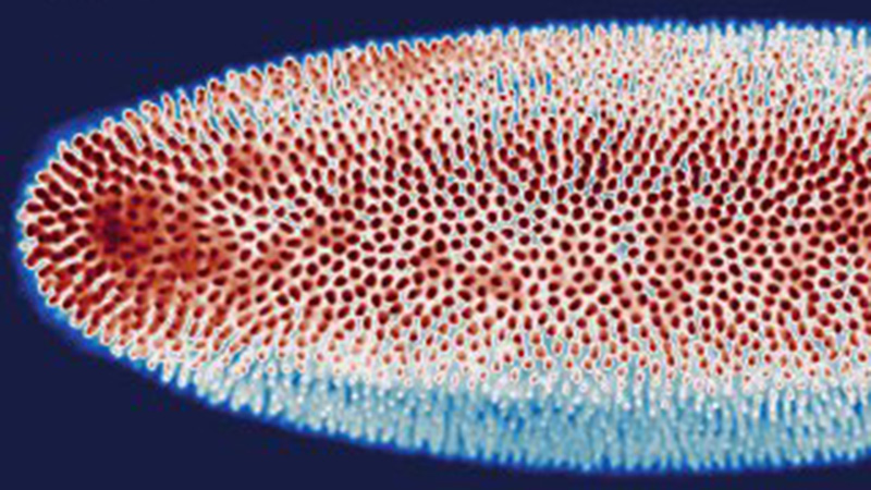
In Vivo Time-Lapse After Photomanipulation in Zebrafish

Time-lapse imaging of zebrafish distal tubules after localized photodamaging can provide novel insights into understanding wound healing processes over time. The design of Papilio5D sample mounting within separate chambers enables imaging under different treatment conditions to be studied at the same time. This allows comparative insights into how pharmaceutical components might impact wound healing.

In this example, regeneration of the distal tubule is studied in the embryonic zebrafish kidney using the cdh17:egfp transgenic line after photodamaging with a photomanipulation module.

Time-lapse imaging of the zebrafish distal tubule after PM.

Samples provided by Leon Rapp (Westhoff lab). The experiment is part of the INFLANET project (Sankeert Satheesan / Jochen Gehrig, Luxendo) in collaboration with Prof. Jens Westhoff (Children’s University Hospital Heidelberg). Acquisition details: 25x 1.1 NA, 242 slices at 1μm, every 10 min for 10 hours, Gauss 1.6μm, 2x1 tiling, Fused view.

Specifications

Select Papilio5D Technical Specifications

Objective Tube Lens Magnification Pixel Size [nm] FOV [µm]
Detection 2x Nikon CFI APO LWD 25x 1.10 NA 100 12.5x 520 1065
  200 25x 260 532
  300 37.5x 173 353
  400 50x 130 266
Detection 2x Nikon CFI APO LWD 16x 0.8 NA 100 8x 813 1664
  200 16x 406 832
  300 24x 271 555
  400 32x 203 416
DOWNLOAD THE BROCHURE FOR MORE INFORMATION

Illumination Optics

  • Chromatic correction from 440 to 660 nm
  • Light-sheet generation by Gaussian or Bessel beam generation and beam scanning
  • Flexible light-sheet thickness (2 µm to 6 µm)
  • Motorised ND filter set (90%, 10%, 1%) for high flexibility in using low laser power
  • Thorlabs 20 W 0.6 NA water immersion objective lens

Detection Optics

  • Two Nikon CFI Apo 25x W 1.1 NA water immersion objective lens or two Nikon 16x W 0.8 NA water immersion objective
  • Two spectral detection channels, each equipped with a fast filter wheel (10 positions)
  • Filters adapted to the selected laser lines
  • Three motorised dichroic mirror positions for simultaneous dual channel acquisition (optional)
  • Motorised collar ring control (25x)
  • Two high-speed sCMOS cameras Hamamatsu Orca Flash 4.0 V3 (or others)
  • Maximum frame rate >80 fps at full frame (2048 × 2048 pixels of 6.5 µm × 6.5 µm size) and up to 500 fps at subframe cropping
  • Peak quantum efficiency (QE): 82% @ 560 nm

System Comparison
* COMPATIBLE MODULE

  Papilio5D
Dual-view
MuVi SPIM
Multiview
InVi SPIM Lattice Pro
Inverted View
LCS SPIM
Large Cleared Samples
TruLive3D Imager
Benchtop Design
Configuration
Horizontal – Dual Detection Horizontal Multiple-View
Inverted
Inverted - Dual Illumination
# Lenses

3 lenses (1 IO, 2 DO) 4 lenses (2 IO, 2 DO)
2 lenses (1 IO, 1 DO) 3 lenses (2 IO, 1 DO)  
Multi-View
       
Live/Fixed Samples
 
Cleared Samples
     
Best Embedding
Sample holder/three dishes/ two separate chambers Capillary/FEP tube w/ agarose 3D stage
FEP foil; glass slides
Quartz-crystal cuvette TruLive3D dishes and FEP foil
Photomanipulation*
 
Environmental Control*  
Destriping/Uniform Illumination* ✓ (w/ Advanced Illumination Module)
Software

All-in-One LuxBundle Software

Intuitive design

Bruker's LuxBundle software saves time and enhances productivity by providing:

  • All-in-one, easy-to-use interface for acquisition, viewing, and post-processing
  • Fully scriptable microscope control and post-processing via open interface (e.g., Python or any other language), ready for custom "smart" microscopy
  • High reproducibility of experiments: all parameters are saved in the metadata and configurations can be saved for future experiments
  • Data formats (.tiff, .hdf5, .ims) compatible with common image processing software: Imaris, Aivia, BigDataViewer, Arivis, Fiji, Python, Matlab, Napari


Impressive Image Post Processor

Our dual-view light-sheet microscopes record a sample from different angles/views and generate images composed of multiple tiles. The LuxBundle software ensures high-quality, 360° crisp images of the sample that compensate for absorption and scattering. Features include:

  • Multi-color alignment
  • Tile stitching of hundreds of tiles for large samples
  • Multi-view image fusion and deconvolution


3D Data Viewer

LuxBundle's integrated 3D data viewer allows researchers to inspect the entire dataset directly after acquisition. This gives users control over their data with key capabilities, including:

  • The ability to turn tile stitching on or off
  • Both raw and post-processed images
  • Fast viewing of multi-terabyte data sets
  • Flexible options to draw and annotate regions and landmarks
Cleared mouse embryo labeled with methylene blue (cyan) and showing autofluorescence (magenta). Image composed of 12 tiles and 920 planes, all processed and stitched with LuxBundle (Image courtesy of Montserrat Coll Lladó, European Molecular Biology Laboratory, Barcelona, Spain)
Papilio5D FAQs

Frequently Asked Questions

What sample types can be imaged with Papilio5D?

Papilio5D is designed for high-sensitivity imaging of organoids, 3D cell cultures, zebrafish larvae, and other optically dense and delicate biological samples.

How many samples can Papilio5D image simultaneously?

The sample holder supports three dishes with two chambers each, allowing imaging of six experimental conditions in parallel.

What detection objectives and magnifications are available?

Papilio5D supports Nikon CFI APO LWD 25x 1.10 NA and 16x 0.8 NA objectives. Magnification ranges from 8x to 50x, depending on tube lens configuration (100–400 mm).

What beam types and illumination modes are supported?

Papilio5D supports Gaussian and Bessel beam illumination. Light-sheet thickness is adjustable from 1 µm to 6 µm.

What acquisition modes are available?

Papilio5D offers area mode for full-field acquisition and line mode with rolling shutter for improved contrast in thick or scattering samples.

What environmental controls are included?

Papilio5D provides temperature control (20–34°C), gas regulation (CO₂ and O₂), and humidity management for long-term imaging.

What are Papilio5D’s pixel size and field of view specifications?

Pixel sizes range from 813 nm to 130 nm. Field of view varies from 1664 µm (at 8x) to 266 µm (at 50x).

What are the system’s frame rate and quantum efficiency?

Frame rates exceed 80 fps at full frame and reach up to 500 fps with subframe cropping. Peak quantum efficiency is 82% at 560 nm.

How can I request more information about this system or determine whether this system is the best fit for my needs?

Contact us to request more information or discuss your application with a light-sheet microscope expert. You can also download the brochurerequest a quote or see a side-by-side comparison of our light-sheet microscope systems.

Contact Expert

Contact Us About Papilio5D

Input value is invalid.

* Please fill out the mandatory fields.

Please enter your first name
Please enter your last name
Please enter your e-mail address
Please enter your Company/Institution


Please enter a valid phone number

ⓘ Used to provide a faster response to your request/question.

Please enter a valid phone number

ⓘ Used to provide a faster response to your request/question.

Please enter a valid phone number

ⓘ Used to provide a faster response to your request/question.

Please enter a valid phone number

ⓘ Used to provide a faster response to your request/question.

Please add me to your email subscription list so I can receive webinar invitations, product announcements and events near me.
Please accept the Terms and Conditions

             Privacy Notice   Terms of Use