Combining Electron Paramagnetic Resonance and Atomic Force Microscopy to Elucidate Oxidative Stress in Eye Disorders
Oxidative damage is thought to underlie many age-related disorders of the eye, and can be caused by oxidative molecules as well as photooxidation from UV light. The risk of AMD increases with exposure to light, highlighting the detrimental potential of oxidation to the aging eye.
The most relevant sources of oxidative stress to the eye are not completely understood, and future therapeutic interventions will require a robust understanding of risk factors and causal mechanisms. Anti-oxidants are known to be protective in AMD, however, the development of safer and more efficacious agents could be possible with a better understanding of the relevant biology.
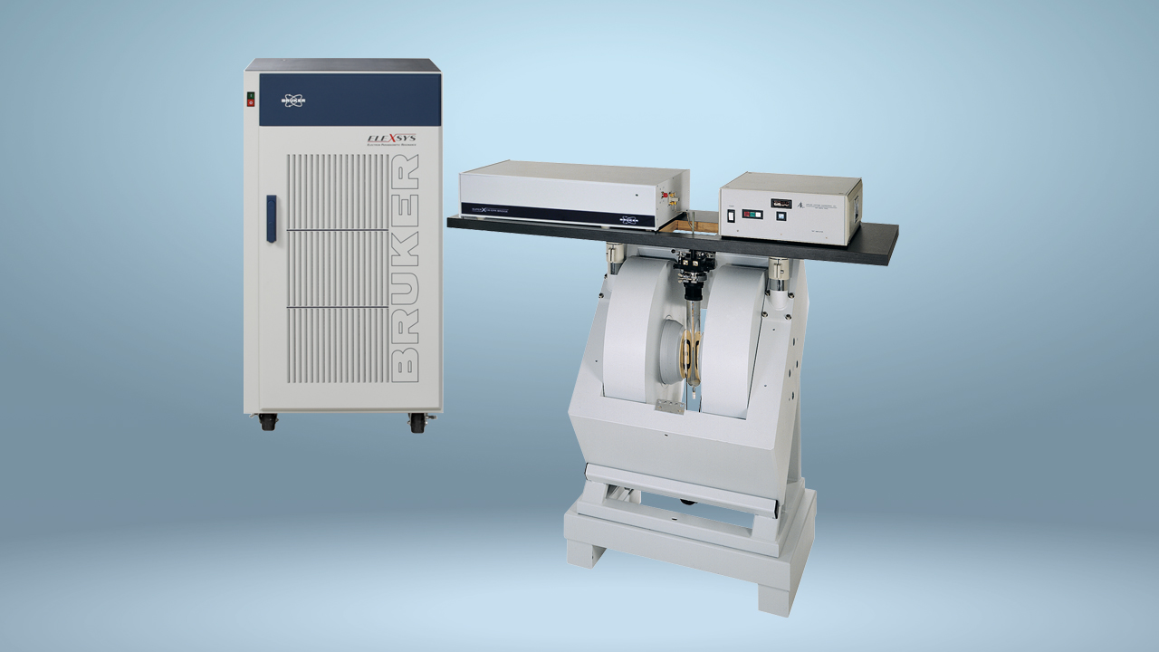
In order to better understand factors influencing age-related oxidative damage, a recent study evaluated the role of metal ions in accelerating oxidation in melanin-containing melanosomes from bovine eyes. Dr. Andrzej Zadlo and colleagues at Jagiellonian University in Krakow, Poland performed electron paramagnetic resonance (EPR) spectrometry, EPR oximetry and atomic force microscopy (AFM) using Bruker instruments. The combination of EPR and AFM allowed the researchers to determine the dynamic changes induced in melanin’s structure following oxidative stress, which may be relevant to various human disorders of the eye including AMD.
Published in April of 2017 in the journal Cell Biochemistry and Biophysics, their paper “Redox Active Transition Metal Ions Make Melanin Susceptible to Chemical Degradation Induced by Organic Peroxide,” shows that redox active metal ions, especially iron, accelerate oxidative changes in melanin induced by tert-butyl hydroperoxide (TBH), a known oxidizing agent.
The authors found that TBH alone induced only modest oxidative changes in melanin, as determined by EPR. However, when bovine melanosomes were co-incubated with TBH alongside metal ions, the presence of iron and copper ions was associated with elevated degradation of melanin. Zinc had no effect on melanin oxidation, indicating that this effect may be limited to redox active metal ions in the eye.
AFM analysis showed that melanosomes degraded by oxidation had reduced surface roughness, and melanin switched from an anti-oxidant to a pro-oxidant. Conversely, the authors found no change in oxidation induced by UV light when any of the tested metal ions was added. These results are physiologically relevant, as iron is implicated in various forms of eye disease, and increased iron levels have been observed in age-related eye disorders.
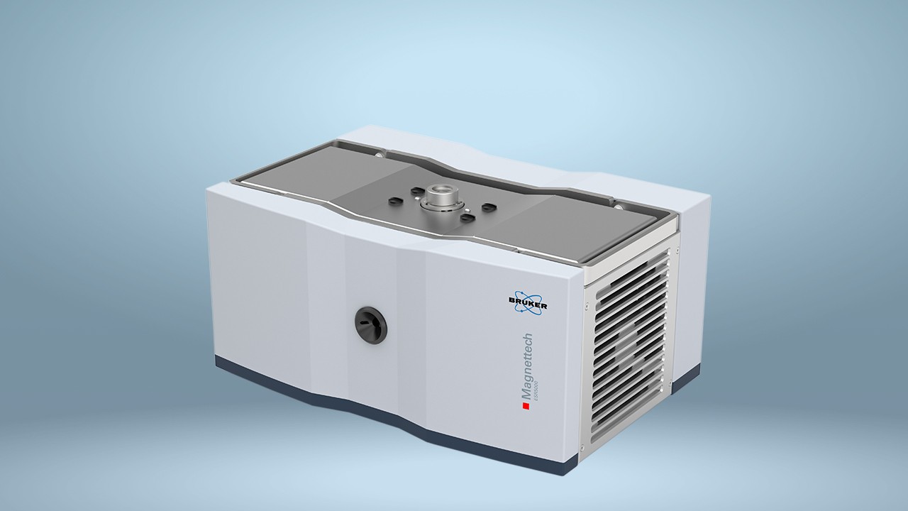
This study highlights the power of EPR and AFM technologies, and the Bruker equipment used to perform it, for studies seeking to understand complex structure-function relationships of proteins. The Bruker EMX-AA EPR allowed the researchers to determine the oxidation and degradation patterns of melanin in the presence of lipids and metal ions in the context of the aging eye.
By using both spectrometry and oximetry, Dr. Zadlo and colleagues were able to determine the relative contributions of molecular oxidizing agents and photooxidation to melanin degradation. This may provide useful information to researchers seeking to develop therapeutic intervention These data indicate that pharmacological agents aimed at reducing oxidative damage may provide greater benefit than increased use of sunglasses or avoidance of direct sunlight. In addition, the images obtained with the Bruker Bio-Scope Catalyst AFM gave the authors a unique perspective on the surface structure of the melanosomes.
With EPR spectroscopy unpaired electrons, such as oxygen radicals, can be tracked dynamically to offer insights into the structure and stability of proteins. EPR offers improved sensitivity, and allows researchers to perform experiments on larger molecules and within shorter time-frames, when compared with nuclear magnetic resonance (NMR).
AFM uses a scanning probe to determine the 3-dimensional structure of molecules, differing from scanning electron microscopy (SEM), which uses a beam of highly energized electrons to generate an image. AFM offers significant advantages over SEM, particularly on samples with smooth surfaces or those that must be immersed in liquid during imaging.
Bruker, the world’s premier supplier of EPR and AFM systems, offers highly sensitive and accurate spectrometers and microscopes for use in fields as diverse as structural biology and quantum physics.
References:
- Klein R, Chou CF, Klein BE, et al. Prevalence of age-related macular degeneration in the US population. Arch Ophthalmol. 2011;129:75-80.
- Hammond BR, Johnson BA, and George ER. Oxidative Photodegradation of Ocular Tissues: Beneficial Effects of Filtering and Exogenous Antioxidants. Exp Eye Res. 2014;129:135-50.
- Köberlein J, Beifus K, Schaffert C, et al. The economic burden of visual impairment and blindness: a systematic review. BMJ Open. 2013;3:1-14.
- Sui GY, Liu GC, Liu GY, et al. Is sunlight exposure a risk factor f or age-related macular degeneration? A systematic review and meta-analysis. Br J Ophthalmol. 2013;97:389-394.
- National Eye Institute. NIH study provides clarity on supplements for protection against blinding eye disease. https://www.nei.nih.gov/news/pressreleases/050513.