Sedimentology & Stratigraphy with the M4 TORNADO
Why Use the M4 TORNADO for the Characterization of Sedimentary Rocks?
Bruker’s M4 TORNADO micro-XRF spectrometer provides minimally invasive, rapid spatial elemental characterization of a wide range of sedimentary materials from unconsolidated or wet sediments (lacustrine or marine) to sedimentary rocks, as cut samples or drill core.
With a spot size resolution to below 20 microns and ability to simultaneously detect major, minor and trace elements, characterization of fine features such as the chemical variation in laminated sediments is easily achieved. With full quantification capability, data may be correlated with other bulk techniques to build detailed and robust chemo-stratigraphic models.

Compositional Characterization of Laminated Sediments
Marine and lacustrine sediments commonly preserve compositional variations that are a reflection of cyclic depositional events and climate variations. The sediments may also locally record evidence for catastrophic events impacting a location, all of which are targets for scientists to better understand earth events on local, regional, and global scales. One key to unraveling these event histories is accurate and robust characterization on scales similar to those variations preserved in the samples.
The small spot size and high element resolution achieved with ease using the M4 TORNADO micro-XRF spectrometer makes the instrument ideal for characterization of finely laminated sediments. Complementary to traditional single-line scanning XRF, the M4 TORNADO provides a more comprehensive and detailed picture of vertical and lateral variations in a sedimentary core, or other samples.
Learn more about the study here:
- Cornard, P.H., Degenhart, G., Tropper, P., Moernaut, J., Strasser, M. (2023). Application of micro-CT to resolve textural properties and assess primary sedimentary structures of deep-marine sandstones. The Depositional Record – The Journal of the International Association of Sedimentologists, 10, 559-580.

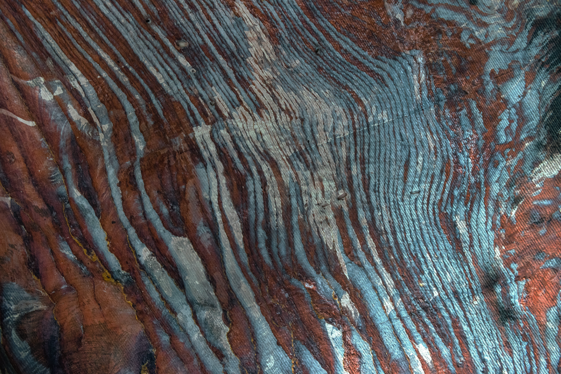
Elemental Variation in Banded Iron Formations
Banded Iron Formations (BIFs) are both records of an important part of Earth’s history and the largest source of iron ore mined worldwide. Formed when early cyanobacteria began to produce enough oxygen to react with dissolved iron in the world’s oceans causing iron oxide minerals to precipitate and be deposited on the ocean floors. Formed between ~3.8 and 1.7 billion years ago, BIFs are only preserved in a few locations worldwide but are keys to understanding processes operating in the early oceans.
Micro-XRF provides a ‘bigger picture’ of any BIF sample than can viewed based on traditional thin sections alone, while still retaining a small (<20 um) spot size and with the benefit of sensitivity to trace levels. Elemental mapping by the M4 TORNADO reveals deep levels of detail in any sample, allowing a researcher to understand the chemical complexity that may be preserved in such samples.

Full Spectrum Provenance Analysis of Unconsolidated Sediment and Crushed Samples
Understanding the source of components in a sedimentary rock is a key component of studies about sedimentary processes and terrane evolution. Information such as mineral species, abundance, and grain sizes, are commonly targeted using laborious optical microscopy techniques, or through analysis by scanning electron microscope. In some cases, samples are screened for dateable minerals, especially zircon, to determine the ages of rocks forming the source materials to a sediment and also the tectonic setting of erosion and deposition.
Traditional techniques typically require extensive sample preparation, such as thin sections or polished epoxy mounts. In contrast, the M4 TORNADO micro-XRF spectrometer allows rapid screening of particulate samples to identify the presence of even a trace abundance of minerals. Elemental mapping may be conducted on non-flat samples, including scatter-mounts of particulates without the need for mounting in epoxy, polishing, or carbon coating, making quick screening simple and easy to achieve. The added benefit of micro-XRF, as an ED-XRF technique, is that an entire energy spectrum is collected simultaneously at each point, allowing comprehensive characterization of a sample and detection of minerals not expected within a sample, and a robust full sample provenance in a much-reduced period of time in a systematic manner.

See the Data and Images in More Detail


Interested in Transforming Your Lab with the Power of the M4 TORNADO?
Whether you're exploring new instrumentation, planning your next grant proposal, or simply curious about how micro-XRF could fit into your workflow - we're here to help.
Explore How the M4 TORNADO Supports Your Research - Let's talk
Connect with our regional team to discuss your geoscience applications. We’ll help align your goals and route next steps to the right Bruker specialists. Book a short, no‑obligation meeting to get started.
Request Pricing
Get a customized quote based on your lab’s needs and configuration preferences.
Request a Live Demo
See the M4 TORNADO in action with real academic use cases and sample workflows.