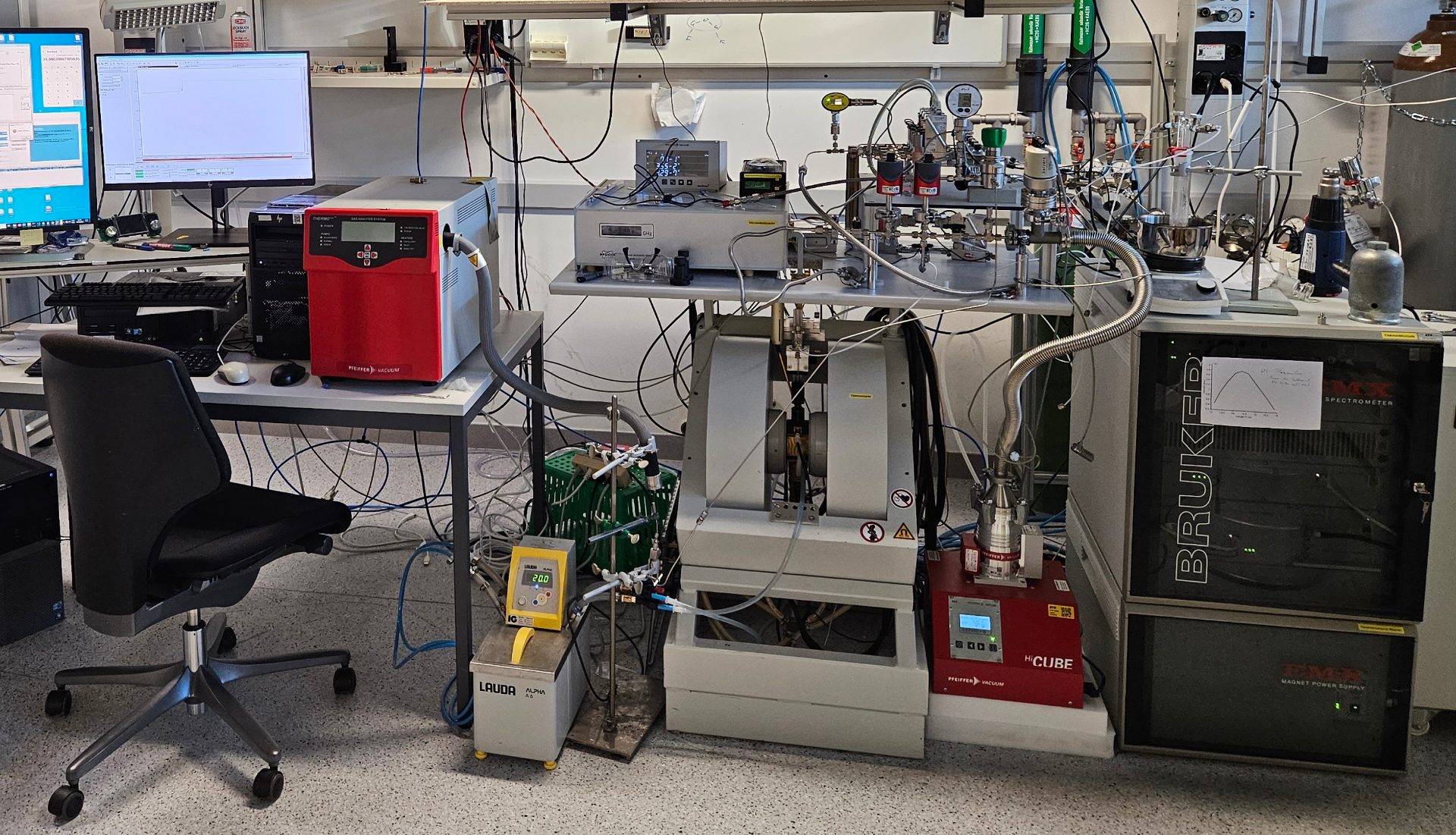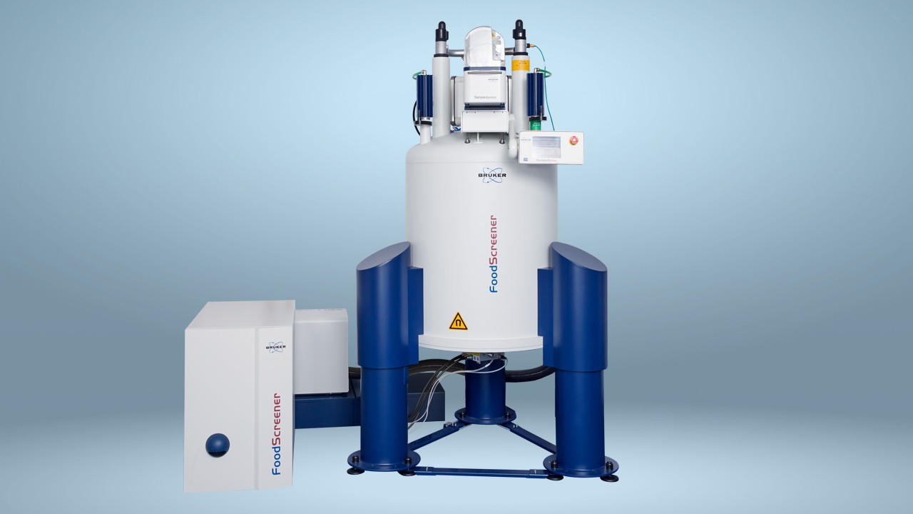

EPR spectroscopy: A unique method for free radical analysis
Electron paramagnetic resonance (EPR) spectroscopy is a method for examining materials with unpaired electrons, such as free radicals, many transition metal and rare earth ions, and paramagnetic defects in solid materials. In fact, it is the only method that directly detects unpaired electrons, – though techniques such as fluorescence can provide indirect evidence of their existence.[1]
Although as a technique it is similar to nuclear magnetic resonance (NMR) spectroscopy, EPR excites the spins of electrons rather than of atomic nuclei and can shed light on the spatial and electronic structure of molecules with unpaired electrons.
In addition, analysis of EPR spectra can provide information on the identity of detected paramagnetic species and their concentration. It is applicable to most samples that feature unpaired electrons,[2] from basic organic compounds to complex biomolecules. It is used for in vivo research in clinical fields,[3] in pharmaceuticals to detect drug impurities,[4] and in material characterization.
“Compared to nuclei, electrons have much larger magnetic moments, which means we can both detect smaller concentrations and look at faster motion,” says Dr. Gunnar Jeschke, full Professor in Electron Paramagnetic Resonance at ETH Zürich. “And while it is less sensitive than optical techniques, it allows us to target the center of reactivity, which is often the unpaired electron, and address the exact question that we want to address – there is no background interference. This also applies in combination with spin labelling and makes EPR unique as a method.”
Research at the molecular level
Dr. Jeschke is based in the Institute of Molecular Physical Science at ETH Zürich, where several teams conduct fundamental research into the properties and matter at atomic and molecular levels. “The institute’s research has implications for the entire range of the natural sciences, from physics to chemistry and materials sciences to biology,” says Dr Jeschke.
Dr. Jeschke heads a team of chemists, physicists, and technical staff who use EPR spectroscopy to understand structure formation on length scales between 0.2 and 10 nanometers. The emphasis is on distance measurements in the nanometer range between spin probes by advanced pulsed techniques and on operando and ex situ characterization of heterogeneous catalysts.
“We use the unpaired electrons mainly of stable free radicals but also of metal ions to get information on the environment,” he says. “One of the main applications is measuring distance distributions in the nanometer range, mostly on proteins, to find out about protein structure and flexibility.”
The research team’s use of continuous-wave and echo-detected EPR is complemented by electron nuclear double resonance (ENDOR) and electron spin echo envelope modulation (ESEEM) techniques that probe the sub-nanometer range by measuring hyperfine couplings to nuclei in the vicinity.
EPR: research and applications
In the presence of a static magnetic field, the energy levels of electron spins are split and EPR spectroscopy detects transitions between them caused by electromagnetic radiation. By analyzing the resulting EPR spectra, valuable information about electron spin states, coordination environments, and molecular dynamics can be obtained.
In the analysis of proteins, EPR offers many advantages over other analytical techniques. In addition to not being constrained by protein size, it can also target biomolecules at concentrations around the micromolar range and does not require crystallization. It also allows the detection of proteins in solution without the need for biomolecules to be isotope-labeled.[5]
According to Dr. Jeschke, the only technique that compete with EPR on measurements of distances in the nanometer range in disordered systems is Förster resonance energy transfer (FRET), which is based on energy transfer between chromophores (light sensitive molecules).
“In the field of protein structure determination, EPR has a unique length scale of between about 20 and 100 angstroms,” he says. “FRET is more sensitive and accesses a similar length scale between 10 to 100 angstrom distances – but EPR gives more accurate information, and most importantly EPR can see the distribution of distances, something that is vitally important for flexible systems.”
Understanding disease-forming proteins
One of the key areas of Dr. Jeschke’s work involves the understanding of ribonucleic acid (RNA)-based proteins, which are involved in the processes of making new proteins in the cell and play an important function in the regulation of gene expression.
“An interesting feature of RNA-binding proteins is that in living cells, they undergo the liquid-liquid phase separation (LLPS) process, leading to the formation of ‘membraneless organelles’,” he says. “If these membraneless organelles do not function in the normal way, they can lead to the development of illnesses such as cancer or neurodegenerative diseases.”
LLPS in vitro leads to formation of protein-rich droplets, and the team has found that continuous-wave EPR of spin-labelled proteins distinguishes between protein molecules inside and outside such droplets. Double electron electron resonance (DEER) is one of the few techniques which can elucidate the structure of proteins in droplets.[6]
“EPR spectroscopy gives unique information,” he says. “Normally, the protein doesn't provide us with a signal, but by spin-labelling a small fraction of the protein, we can pick out isolated molecules within the large volume of a liquid droplet and look at the protein’s structure.” In this way, he hopes to understand how proteins in a liquid droplet interact. “It allows us to monitor the ageing of the droplet to understand a process that can cause disease.”
Making chemistry greener
Dr. Jeschke also conducts research on heterogeneous catalysis, where reactants and products exist in a different phase to the catalyst. Heterogeneous catalysis is used in the majority of industrial chemical processes to produce various chemical compounds such as ammonia and is regarded as a key method for reducing energy consumption in the production of chemicals and biofuels.[7]
Recently published research[8] by a consortium involving Dr. Jeschke looked at ways to produce methanol with a lower carbon footprint. EPR spectroscopy allowed Dr. Jeschke and the team to ascertain that zinc-zirconia (ZnZrOx) – a more cost-effective and sustainable material – had potential as a catalytic material in methanol production from CO2.
The overall goal of the heterogeneous catalyst research is to find more environmentally friendly ways of making chemicals and helping the chemistry industry to become more sustainable. “Our aim is to make chemistry greener; to find catalysts that can create the products that we need in a way that is much less harmful to the environment,” he says.
Benefit to society
Dr. Jeschke is currently working on two papers related to proteins undergoing LLPS.
“We are discovering how the binding of RNA changes the shape and the structure of the protein, and this gives some insight into why the protein can bind different types of RNA – one of its functions in the cell,” he says. The second paper focuses on a protein called FUS, which is related to Amyotrophic Lateral Syndrome (ALS). “We have discovered that when this protein goes through LLPS, the basics of this LLPS can be understood in rather basic polymer physics terms – something which has not previously been clear, and which is a rare finding in biology.”
He and his researchers also plan to take their protein studies further in the near future. “We do not only want to know the structure of a single protein, but we also want to know how they are related to each other,” he says. “We are already conceiving ideas as to how we do the experiments for this, though I suspect it will be a few years from now before have the first results.”
Dr. Jeschke says that his work in structural biology and in heterogeneous catalysis will be used in practical application.
“In heterogeneous catalysis, it will lead to more energy efficient and less environmentally harmful ways of making chemicals,” he says. “And, ultimately, in biology, it will give us a better understanding of cellular processes, in turn leading to the development of drugs that help to address under-researched health problems.”
The team may be conducting fundamental research, but Dr. Jeschke believes very firmly that the benefits are wide-reaching. “The societal benefit of fundamental research is, of course, that we satisfy society’s curiosity,” he says. “But without continuous investment in new fundamental research, applied research would die out very quickly. Exploring the capabilities of EPR spectroscopic research is crucial for wider applications that help solve some of the big picture issues humanity is facing.”
About Dr. Gunnar Jeschke
Dr. Gunnar Jeschke has been full Professor in Electron Paramagnetic Resonance at ETH Zürich since April 2008. His electron paramagnetic resonance research incorporates elements of molecular and polymer physics, transition metal chemistry, and molecular biology. He contributes to understanding of spin dynamics, developments in EPR instrumentation and pulse sequences for hyperfine spectroscopy and distance distribution measurements, integrative structural biology, and ensemble modelling and analysis of proteins and their complexes with RNA.
About Bruker Corporation
Bruker is enabling scientists to make breakthrough discoveries and develop new applications that improve the quality of human life. Bruker’s high-performance scientific instruments and high-value analytical and diagnostic solutions enable scientists to explore life and materials at molecular, cellular, and microscopic levels. In close cooperation with our customers, Bruker is enabling innovation, improved productivity, and customer success in life science molecular research, in applied and pharma applications, in microscopy and nanoanalysis, and in industrial applications, as well as in cell biology, preclinical imaging, clinical phenomics and proteomics research and clinical microbiology.
References
- Bruker. What is EPR? Available at: https://www.bruker.com/en/products-and-solutions/mr/epr-instruments/what-is-epr/What-Is-EPR.html Accessed Oct 2nd, 2023.
- Wilder, A. EPR: Application. Available at: https://chem.libretexts.org/Bookshelves/Physical_and_Theoretical_Chemistry_Textbook_Maps/Supplemental_Modules_(Physical_and_Theoretical_Chemistry)/Spectroscopy/Magnetic_Resonance_Spectroscopies/Electron_Paramagnetic_Resonance/EPR%3A_Application#:~:text=member%20Alex%20Gunn.-,Experimental%20Applications,complex%20biomolecules%20such%20as%20proteins Accessed Oct 2nd, 2023.
- Schwarz HM et al. Clinical applications of EPR: overview and perspectives. NMR Biomed. 2004: Aug; 17(5): 335-51. DOI: https://doi.org/10.1002/nbm.911
- Bruker. Pharmaceutical applications of EPR spectroscopy. Available at: https://www.bruker.com/en/news-and-events/webinars/2018/pharmaceutical-applications-of-epr-spectroscopy.html Accessed Oct 2nd, 2023.
- Hofmann L & Ruthstein S. EPR spectroscopy provides new insights into complex biological reaction mechanisms. J Phys Chem B. 2022: Oct 6; 126(39): 7486-7494. DOI: https://doi.org/10.1021/acs.jpcb.2c05235
- Ritsch et al. Phase separation of heterogeneous nuclear ribonucleoprotein A1 upon specific RNA-binding observed by magnetic resonance. Angew Chem Int Ed. 2022; 61. DOI: https://doi.org/10.1002.anie.202204311
- Liu Y et al. Heterogeneous catalysis for green chemistry based on nanocrystals. Natl Sci Rev. 2015; 2(2): 150-166. DOI: https://doi.org/10.1093/nsr/nwv014
- Araujo TP et al. Design of flame-made ZnZrOx catalysts for sustainable methanol synthesis from CO2. Adv Energy Mater, 2023; 13, 22o4122. DOI: https://doi.org/10.1002/aenm.202204122


