

How NMR spectroscopy is providing novel insights into
protein structures, dynamics
and interactions
Exploring the molecular world of proteins
Studying protein structure, dynamics, and interactions is pivotal for understanding disease mechanisms and to advance drug discovery and development. By deciphering the three-dimensional shapes and dynamic behaviors of proteins, researchers can pinpoint structural anomalies or alterations in disease-related proteins, shedding light on the molecular underpinnings of various disorders. This knowledge, in turn, aids in the design of targeted therapies and drugs that modulate dysfunctional protein interactions and restore normal protein function.
There are multiple biophysical techniques for analyzing and determining protein structures, each of which has its pros and cons.(1) X-ray crystallography is often used to determine the three-dimensional atomic structure of proteins by analyzing the diffraction patterns of X-rays passed through a crystal lattice. Prof. Polenova comments: “Because proteins are inherently dynamic molecules that can adopt multiple conformations, it makes it difficult to capture all of these structural states using this method alone. Also, obtaining high-quality protein crystals suitable for X-ray crystallography can be laborious and sometimes impossible for certain proteins.”
Cryo-electron microscopy (cryo-EM) is another technique that has significantly contributed to protein structure determination. It is a powerful method that allows the visualization of biological macromolecules and structures at near-atomic resolution by freezing samples in vitreous ice and using electron microscopy to capture their detailed images. (2)
“Although cryo-EM provides high-resolution images, like X-ray crystallography, it encounters challenges when dealing with intrinsically disordered proteins or proteins lacking well-defined three-dimensional structures,” Prof. Polenova explains. “This is because disordered proteins lack the stable structures that cryo-EM traditionally relies upon for capturing high-resolution images, making it difficult to obtain structural data as disordered proteins appear faint and blurry in images or the disordered regions are missing altogether.”
An integrated analytical approach
Prof. Polenova adopts an integrated approach, by combining various experimental and computational techniques to achieve a comprehensive understanding of how protein structures affect their biological functions.
The primary analytical technique used is nuclear magnetic resonance (NMR) spectroscopy, particularly solid-state magic angle spinning (MAS) NMR, which provides atomic-resolution protein structure information. NMR is a non-destructive analytical tool that uses the inherent magnetic properties of specific atomic nuclei to reveal the structure, identity, concentration, and behavior of molecules. It can be performed both on samples in solution (solution NMR) or on insoluble proteins and protein assemblies that are prepared either as microcrystals, or sedimented or lyophilized samples (solid-state NMR). (3)
“MAS NMR involves rotating the sample at a specific angle to achieve high spectral resolution, for non-liquid samples. This technology enables us to examine both static structures and conformational dynamics, and to unveil structural details of proteins and nucleic acids that remain “invisible” to other analytical techniques because of dynamic disorder,” Prof. Polenova explains. “For example, NMR, both solution and MAS, allows for studies of intrinsically disordered proteins and monitoring biochemical reactions in real-time, providing unique valuable insights which would otherwise not be possible to obtain,” she continues.
Utilizing NMR to understand virus protein assemblies
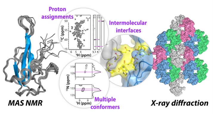
One branch of Prof. Polenova’s research is to examine SARS-CoV-2 structural proteins, which organize the virus's genetic material and help it replicate and cause disease. The nucleocapsid (N) protein is one of these structural proteins. Scientists have long been interested in a specific part of this protein called the N-terminal domain (NNTD) because it interacts with the virus' genetic material and could be a target for drugs or vaccines.(4)
“To understand this protein better, we used solid-state MAS NMR and X-ray diffraction to study its atomic structure.(5) This combined approach helped us see how the atoms are arranged in this domain of the protein and how it interacts with other molecules – information that would not be attainable without NMR. Gaining insights into the protein structures in this way is essential as it offers guidance in designing treatments to fight the SARS-CoV-2 virus,” states Prof. Polenova.
Another branch of her virus protein research focuses on HIV protein assemblies. Human immunodeficiency virus (HIV) is the causative agent for acquired immune deficiency syndrome (AIDS), which has taken the lives of more than 40 million people across the globe to date.(6) Scientists are continually searching for effective treatments to prevent HIV from progressing to AIDS.
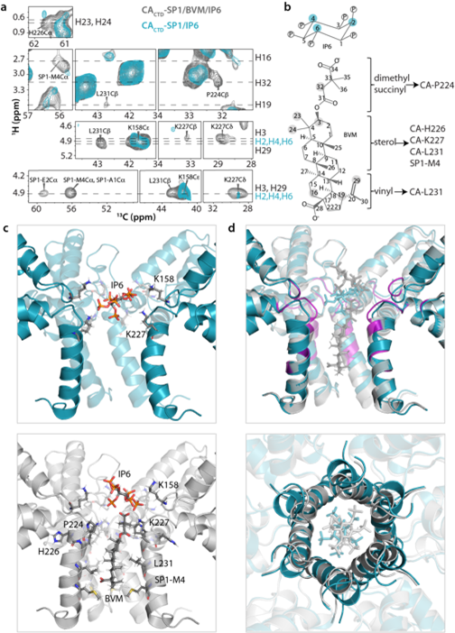
At the forefront of this ground-breaking research is the Pittsburgh Center of HIV Protein Interactions (PCHPI), a truly interdisciplinary team, established in ca. 2007 by Prof. Angela M. Gronenborn, which Prof. Polenova and her laboratory are part of. In collaboration with multiple PCHPI scientists, Prof. Polenova’s team made an important discovery in 2017 – they found that molecular dynamics play key roles in HIV maturation, suggesting potential ways to interfere with the HIV infection.(7) Prof. Polenova explains that the HIV-1 virus goes through a complex maturation process before it becomes infectious: “As it develops, protein building blocks are cleaved from the master protein called Gag. But the final step in the virus’ evolution to becoming infectious had long remained a mystery, particularly the role of the final-step maturation intermediate protein, where a 14-residue spacer peptide 1 is uncleaved from the capsid protein (CA-SP1).”
Using a combination of techniques, including MAS NMR, cryo-EM, and computer simulations, the PCHPI team found that this SP1 peptide needed to be mobile for the cleaving process to be effective. She says: “Viruses like HIV and their constituent proteins together with nucleic acid molecules are dynamic entities. The SP1 peptide is always there in the final maturation intermediate before being cleaved off by the viral protease, but we were surprised that it is so dynamic. Small-molecule maturation inhibitors, such as Bevirimat (BVM), act by binding inside the pore formed by 6 SP1 peptides in the assembly and arresting the SP1 dynamics.(8) Without the help of MAS NMR techniques we would not be able to see the dynamic SP1 peptide and the structure of BVM-bound CACTD-SP1 assembly.”

About Prof. Tatyana Polenova
Tatyana Polenova is a Professor of Chemistry and Biochemistry at the University of Delaware and leads a team of scientists at the Polenova Laboratory. Her research focuses on investigating the structure and dynamics of biological systems and complex formulations in the solid state using NMR spectroscopy, combined with computational, and biochemical methods. Alongside her team at the Polenova Laboratory, she is currently studying HIV-1 capsid and SARS-CoV-2 protein assemblies, microtubule- and actin- associated protein assemblies, and vanadium-containing haloperoxidases, with the aim of better understanding disease mechanisms and assisting with drug discovery and development, as well as biotechnology applications. Prof. Polenova serves as the Editor in Chief of the Journal of Magnetic Resonance and a Co-Director of the Pittsburgh Center for HIV Protein Interactions.
About the Polenova Laboratory
The research at the Polenova Laboratory primarily focuses on the development and utilization of innovative magic angle spinning nuclear magnetic resonance (MAS NMR) and dynamic nuclear polarization (DNP) enhanced MAS NMR spectroscopy techniques to investigate structures, dynamics, and functions of biological assemblies, small-molecule pharmaceuticals and biologics, and proteins in cellular environments. The lab is particularly interested in studying systems where MAS NMR can provide unique insights that are often unattainable through other methods. This includes the study of microtubule- and actin- associated protein assemblies with relevance to various diseases, HIV-1 viral protein assemblies, SARS-CoV-2 nucleocapsid, biotechnologically significant vanadium haloperoxidases, as well as lipid nanoparticles for drug delivery.
These studies by Prof. Polenova and the PCHPI team are uncovering the structural basis of HIV-1 maturation BVM binding and activity, and providing guidance for the design of more potent inhibitors that could interfere with the HIV maturation and prevent the virus from turning infectious. These improved maturation inhibitors under development represent a novel class of drugs for combating HIV/AIDS that could save lives across the globe.
Investigating cytoskeleton protein assemblies
Prof. Polenova explains that another class of systems she is working on are cytoskeleton protein assemblies, specifically proteins that bind to microtubules and actin. Microtubules are cytoskeleton filaments that play essential roles in maintaining cell structure, organelle transport, cell motility, and cell division. Cells need to organize these microtubules in a specific way for various cellular functions, and they use microtubule-associated proteins (MAPs) to do this.
MAPs control how microtubules form, how stable they are, how they move, and how they interact with the cytoskeleton and other cell parts.(9) “To understand how these proteins work, we need to know exactly how they attach and interact with microtubules at the atomic level. This knowledge about how their structure relates to their function could help us understand diseases better and develop treatments, but we don't have much of this detailed structural information yet,” says Prof. Polenova.
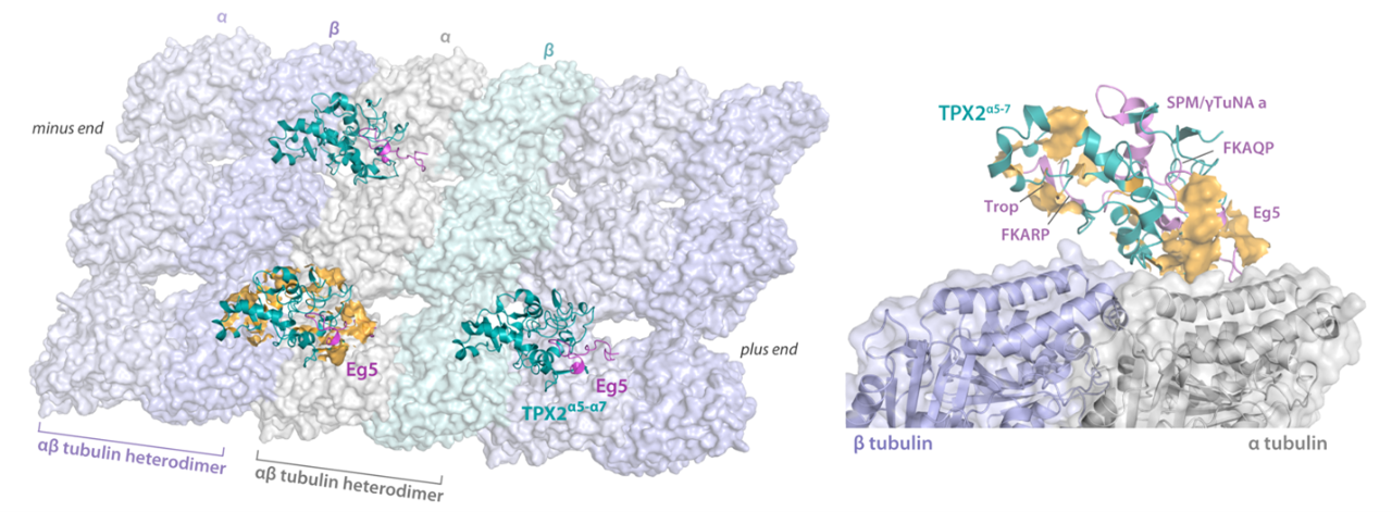
She continues: “Using MAS NMR, we have been able to determine structures of several proteins bound to polymeric microtubules, such as motor protein domain of kinesin-1,(10) and TPX2.(11) These are key MAPs, and TPX2 is intractable by other techniques because of its intrinsic disorder. It is known from prior investigations that, if the function or expression of TPX2 become disrupted, it can lead to genomic instability and affect important microtubule cell division processes.(12) This in turn can give rise to certain human cancers, which means TPX2 may represent an important biomarker for malignancies that can help us catch and treat cancer earlier and more effectively.”
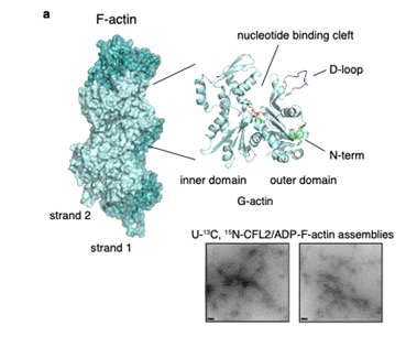
Similarly, actin filaments form part of the cytoskeleton. These filaments undergo polymerization regulated by actin-binding proteins, a process that is essential for various cellular functions. The cofilin family of proteins are regulators of actin severing and filament disassembly, however, the structural basis for this activity is poorly understood, as it was challenging to obtain atomic-level structures of cofilin-actin complexes by traditional structural biology techniques.(13)
Prof. Polenova adds: “Disruptions to actin dynamics can give rise to neurodegenerative diseases, multiple types of cancer, and cardiomyopathy. MAS NMR enables us to observe the atomic-resolution structure of actin-associated proteins involved in this process bound to actin filaments so that we can better understand the mechanisms behind what causes these diseases to arise. Previous research has used cryo-EM to describe the changes in actin filament architecture, but MAS NMR provides atomic level resolution so that we can really see the much finer structural details of cofilin when bound to actin.”
Analyzing protein structures within living cells
Prof. Polenova explains her team’s goal is to apply the techniques they have developed on isolated proteins and protein assemblies to investigate these structures in situ: “In our laboratory, we are particularly enthusiastic about pushing the boundaries of sensitivity. Our aim is to enhance MAS NMR methods to such an extent that we can closely examine the structures and movements of proteins within the intricate context of living cells, allowing us to gain insights into how these molecules function in their natural environment.”
She explains that, together with Prof. Gronenborn, they are increasingly exploring how fluorine-19 (19F) NMR spectroscopy, a technique that uses the NMR properties of fluorine nuclei, can also be used to study the structures and movements of proteins. This method can also be used to investigate how certain proteins interact with drugs or inhibitors that could be used as pharmaceutical treatments, Prof. Polenova concludes: “This area of research is particularly exciting to us, and our findings could have hugely beneficial implications for medicine.”
References
(1)Polenova T, Quinn CM, Gronenborn AM, eds. Integrated Structural Biology. ROYAL SOCIETY OF CHEMISTR; 2023.
(2)Chua EYD, Mendez JH, Rapp M, et al. Better, faster, cheaper: Recent advances in Cryo–electron microscopy. Annual Review of Biochemistry. 2022;91(1):1-32. doi:10.1146/annurev-biochem-032620-110705.
(3)Quinn CM, Polenova T. Structural biology of supramolecular assemblies by magic-angle spinning NMR spectroscopy. Quarterly Reviews of Biophysics. 2017;50. doi:10.1017/s0033583516000159.
(4)Bai Z, Cao Y, Liu W, Li J. The SARS-COV-2 nucleocapsid protein and its role in viral structure, biological functions, and a potential target for drug or vaccine mitigation. Viruses. 2021;13(6):1115. doi:10.3390/v13061115.
(5)Sarkar S, Runge B, Russell RW, et al. Atomic-resolution structure of SARS-COV-2 nucleocapsid protein N-terminal domain. Journal of the American Chemical Society. 2022;144(23):10543-10555. doi:10.1021/jacs.2c03320.
(6)Global HIV & AIDS statistics - Fact sheet. UNAIDS. 2023. Accessed September 19, 2023. https://www.unaids.org/en/resources/fact-sheet.
(7)Wang, Mingzhang, et al. "Quenching protein dynamics interferes with HIV capsid maturation." Nature Communications 8.1 (2017): 1779.
(8)Sarkar, S., Zadrozny, K.K., Zadorozhnyi, R. et al. Structural basis of HIV-1 maturation inhibitor binding and activity. Nat Commun 14, 1237 (2023). https://doi.org/10.1038/s41467-023-36569-y
(9)Janke, C., Magiera, M.M. The tubulin code and its role in controlling microtubule properties and functions. Nat Rev Mol Cell Biol 21, 307–326 (2020). https://doi.org/10.1038/s41580-020-0214-3.
(10)Zhang, Chunting, et al. "Magic-angle-spinning NMR structure of the kinesin-1 motor domain assembled with microtubules reveals the elusive neck linker orientation." Nature Communications 13.1 (2022): 6795.
(11)Guo, Changmiao, et al. "Structural basis of protein condensation on microtubules underlying branching microtubule nucleation." Nature Communications 14.1 (2023): 3682.
(12)Pérez de Castro I, Malumbres M. Mitotic Stress and Chromosomal Instability in Cancer: The Case for TPX2. Genes Cancer. 2012;3(11-12):721-730. doi:10.1177/1947601912473306.
(13)Kraus, Jodi, et al. "Magic angle spinning NMR structure of human cofilin-2 assembled on actin filaments reveals isoform-specific conformation and binding mode." Nature Communications 13.1 (2022): 2114.