
Bruker at IMSC 2026 - Pioneering 4D Multiomics and Functional Proteomics 2.0
Future-Ready Science. Where Breakthroughs Feel Effortless.
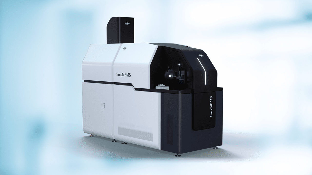
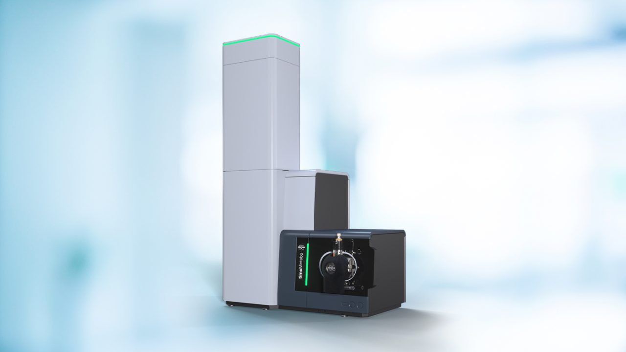
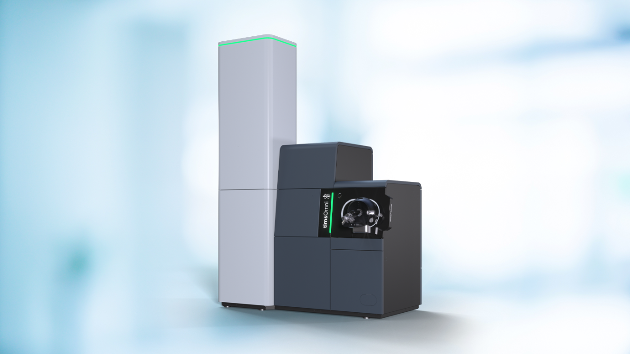
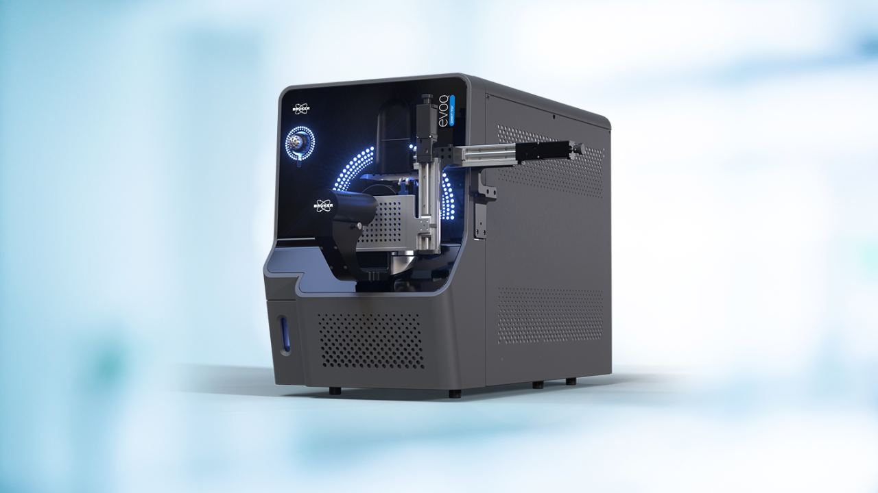
Meet Bruker at IMSC, Centre De Congrés De Lyon, August 22 - 28, and see how next-generation mass spectrometry is transforming 4D multiomics and functional proteomics 2.0. Explore the latest innovations across our portfolio, including timsMRMS, timsOmni™, timsMetabo™, and more.
Visit our team at booth 22 to connect with our experts, learn more about our latest technologies, and see how Bruker solutions are enabling new levels of insight across a wide range of research applications.
Join us throughout the week:
- Lunch Seminar:
“Leveraging TIMS and advanced analyzers to decipher unresolved complexity”
Discover how cutting-edge TIMS-enabled workflows are helping scientists tackle complex molecular challenges. - Lab Talk:
“Faster, Higher, Smaller: The New Pioneers of Mass Spectrometry”
Experience how recent innovations are redefining speed, sensitivity, and resolution in modern MS. - Jochen Franzen Award:
We are proud to sponsor the Jochen Franzen Award for outstanding contributions to innovations in structural, spatial, and separation analysis with mass spectrometry. The award ceremony will take place on August 27, 15:30–16:30.
Whether you are looking for new applications, deeper insights, or inspiration for your research, connect with us at IMSC and be part of the conversation. Details and session information are provided below.

Leveraging TIMS and Advanced Analyzers to Decipher Unresolved Complexity
Join us for this lunchtime seminar and explore new possibilities in molecular analysis: Discover how trapped ion mobility spectrometry (TIMS), combined with advanced mass analyzers and fragmentation technologies, is opening new dimensions in analytical science. This session brings together leading researchers who are applying TIMS-enabled workflows to tackle some of today’s most complex analytical challenges.
From the detailed characterization of highly complex samples such as biofuels, to in-depth analysis of intact proteins, and advanced metabolomics and lipidomics approaches, learn how adding an extra dimension of separation can unlock deeper insights across diverse applications.
Featured presentations
- Pushing the Limits of Environmental Analysis: High-Field FT-ICR Coupled with TIMS for PFAS in Energy and Environmental Samples.
Prof. Carlos Afonso, University of Rouen, France
Explore how TIMS combined with high-field FT-ICR enables the detailed characterization of highly complex environmental samples, including biofuels and PFAS, delivering exceptional resolution, dynamic range, and mass accuracy. - Deep proteoform analysis by ion mobility spectrometry and multi-modal tandem mass spectrometry
Prof. Ole Nørregaard Jensen, University of Southern Denmark, Denmark
Learn how TIMS, integrated with advanced fragmentation technologies and TOF analysis, enables in-depth characterization of intact proteins and proteoforms, providing new insights into protein complexity. - Multidimensional mass spectrometry for metabolomics and lipidomics: Applications in inborn errors of metabolism and research
Dr. Michel van Weeghel, Amsterdam UMC, Netherlands
See how the integration of the collision cross section (CCS) dimension enhances the identification of metabolites and lipids, supporting deeper understanding of disease mechanisms and therapeutic development.
Why attend?
Together, these presentations highlight the power of TIMS-enabled workflows to reveal molecular complexity across environmental and renewable energy research, proteomics, and metabolomics and biomedical applications. Join the session to gain practical insights into how multidimensional separation approaches can elevate your analytical capabilities.
Event details
- Date: Monday, August 24, 2026
- Time: 12:45–13:45 pm CEST
- Location: Room Gratte Ciel 1 & 2, Cité Internationale - Centre de Congrès
Seats are limited - secure your place early to be part of the discussion.
Speakers

Carlos Afonso, Ph.D.
Professor, University of Rouen, Rouen, France

Ole Nørregaard Jensen, Ph.D.
Professor, University of Southern Denmark, Odense, Denmark

Michel van Weeghel, Ph.D.
Research Associate, Amsterdam UMC, Amsterdam, The Netherlands
Faster, Higher, Smaller: The New Pioneers of Mass Spectrometry
Join us and explore how recent advances in mass spectrometry are redefining what is possible. For years, the field was shaped by a fundamental trade-off between speed, sensitivity, and resolution. With the introduction of trapped ion mobility, that limitation has been redefined, opening new pathways to analyze complex biological systems with unprecedented depth and efficiency.
In this 15-minute lab talk, Michael Easterling, Ph.D., will share how scientists are pushing beyond previous boundaries, from reading proteomes at remarkable speed to analyzing intact protein assemblies and enabling insights at the single-cell level. The session highlights how innovation in separation and instrument design is driving a new wave of discovery across life science research.
- Speaker: Michael Easterling, Ph.D. VP Global Imaging, Bruker Scientific LLC
- Date and time: Tuesday, August 25, 14:00–15:00 CEST
- Location: Room Agora, Cité Internationale - Centre de Congrès
Stop by to see how TIMS is redefining the limits of speed, sensitivity, and resolution in mass spectrometry.

Michael Easterling, Ph.D.
VP Global Imaging, Bruker Scientific LLC
Register Now and Secure Your Seat at the Lunch Seminar
Learn how cutting-edge TIMS workflows are driving new discoveries across omics and beyond.
Disclaimer
As you are certainly aware, special compliance regulations apply to public officials* and healthcare professionals** with regard to the event we are planning. If you accept our invitation, we will therefore assume that you will observe the compliance regulations that apply to you and that you have the necessary employer approval.
* Government Official means according the Bruker policies any of the following: any officer, employee or representative of a government (national, regional or local) entity, or any public agency, public authority, department or instrumentality thereof, regardless of their rank or title (e.g. a regulatory official or government inspector); any person working for or advising a government-owned or government-controlled enterprise (e.g. a professor at a government-owned university, or a purchaser at a government-owned hospital); any person working for or advising a national or international non-governmental organization (e.g. an employee of the Red Cross or The World Bank); any person performing a public function or providing a public service, even if that person works for a nongovernmental institution (e.g. private security personnel working in public functions); any person hired to review or accept bids for a government agency; any person with the responsibility to allocate or expend government funds; any person in a public law function, civil servant, judge or military personnel; any person acting for a political party, including party officials, candidates or individuals holding a position in a political party office; members of royal families; or immediate family members of any of the persons listed above. An immediate family member is a grandparent, parent, spouse, significant other, child, or sibling.
** A Healthcare Professional (HCP) is in accordance with the Bruker policies any physician, dentist, nurse, pharmacist or other individual who may prescribe, administer, purchase, dispense, recommend, or supply medical products or treatments or pharmaceutical products. In many cases, Bruker interacts with HCPs who work for state-owned hospitals (e.g. as medical scientists). These individuals will be classified as both HCPs and Government Officials.
For Research Use Only. Not for use in clinical diagnostic procedures.