
AFM in Biosafety Laboratories: Nanoscale Analysis of Biological Agents
Wednesday, December 2 | 8AM PST | 11AM EST | 5PM CET
Delivering Nanoscale Analysis to Biosafety Level Laboratories.
In this webinar, Dr. Sébastien Lyonnais (University of Montpellier) will present his work using AFM in a BSL-3 laboratory to investigate highly infectious viruses. Dr. Human Rezaei (INRAE) will speak on using AFM to study the structural propagation of prions and how non-fibrillar prion oligomers transmit conformational information, and Dr. André Körnig (Bruker) will introduce the new Biosafety Containment Chamber.
Join us for this webinar to learn about:
- Performing AFM in higher biosafety level laboratories
- Using AFM to investigate infectious viruses and prion propagation
- Bruker’s new Biosafety Containment Chamber for AFM imaging and the nanomechanical analysis of biological agents under higher-level biosafety containment conditions.
Wednesday, December 2 | 8AM PST | 11AM EST | 5PM CET
AFM in Biosafety Laboratories: Nanoscale Analysis of Biological Agents
Bringing Atomic Force Microscopy into the BSL-3 Laboratory for the Investigation of Infectious Viruses
Dr. Sébastien Lyonnais, Center for the Study of Infectious Diseases and Anti-Infective Pharmacology (CEMIPAI), French National Centre for Scientific Research (CNRS), France
AFM has proven to be a powerful nanoscale imaging technique for virophysics, enabling high-resolution imaging and nanomechanical mapping of native viruses without labeling under near-physiological conditions. AFM’s advantages are particularly relevant for the study of pathogenic viruses, enabling the analysis of their structure and morphology, the integrity of their membranes and the biophysical changes induced by environmental stress or antiviral treatments. This presentation will introduce the Bruker BioAFM system that we have set up in the biosafety level 3 laboratory at the CEMIPAI platform in Montpellier, and will highlight its applications in exploring the nanoscale world of infectious viruses.
Non-fibrillar prion oligomers transmit folding information
Dr. Human Rezaei, National Institute for Agriculture, Food, and Environment (INRAE), Molecular Virology and Immunology unit, Pathological Macro-assemblies and Prion Pathology Group, France
Prion propagation is traditionally associated with fibrillar assemblies, while the templating capacity of non-fibrillar oligomers remains poorly understood. Here, AFM, combined with rational mutagenesis and hetero-oligomerization assays, reveals how prion protein oligomers transmit folding information. Single-molecule AFM structural analyses identify a modular architecture comprising a globular β-sheet-rich B domain and an elongated E domain. AFM-based characterization of oligomeric assemblies supports a mechanism whereby wild-type PrP recruits assembly-defective variants through structural complementation, initiating conformational conversion within the B domain. These findings establish non-fibrillar oligomers as active conformational templates and alternative platforms for prion structural propagation.
Introducing Bruker’s New Biosafety Containment Chamber
Dr. André Körnig, Senior Application Scientist, Bruker BioAFM
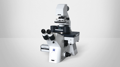
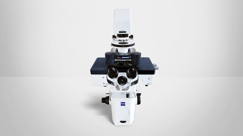
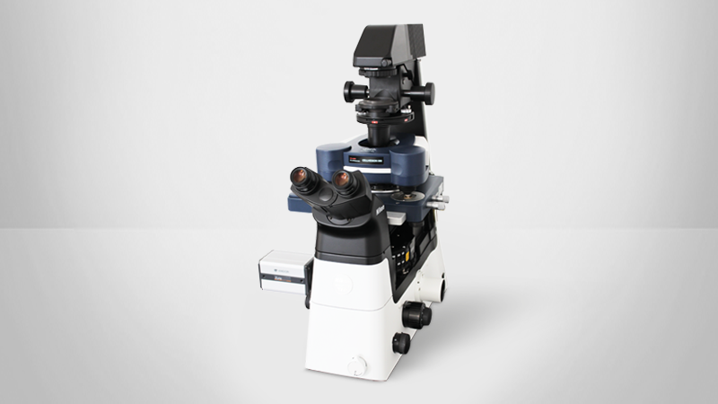
Bruker’s new Biosafety Containment Chamber is a portable, hermetically sealed device with an integrated cantilever holder that enables high-resolution AFM imaging and the nanomechanical characterization of biological agents under higher-level biosafety containment conditions. The chamber supports the AFM analysis of living pathogenic microorganisms under near-physiological conditions and can be integrated with advanced optical microscopes for correlated measurements and multiparametric sample analyses. It is compatible with newer-generation NanoWizard BioAFMs as well as the CellHesion and ForceRobot platforms, extending advanced AFM capabilities to stringent BSL research environments.
Date: December 2, 2026
Time: 8AM PST | 11AM EST | 5PM CET
Location: Online
Featured Technologies:
Contact:
Find out more about the technology featured in this webinar or our other bioafm solutions:
Speakers

Dr. Sébastien Lyonnais, Center for the Study of Infectious Diseases and Anti-Infective Pharmacology (CEMIPAI), French National Centre for Scientific Research (CNRS), France
Dr. Sébastien Lyonnais is Head of the Microscopy Facilities at the high-biosafety laboratory of the CNRS Center for Infectious Diseases CEMIPAI at the University of Montpellier, France. He is a recognized expert in the imaging of viruses, bacteria and human pathogens. He develops and applies BioAFM and fluorescence microscopy technologies in this BSL-3 laboratory for the analysis of infectious biological samples ranging from single viruses to infected tissues. His research is making an important contribution towards a more comprehensive understanding of pathogen structure, dynamics and interactions.

Dr. Human Rezaei, National Institute for Agriculture, Food, and Environment (INRAE), Molecular Virology and Immunology unit, Pathological Macro-assemblies and Prion Pathology Group, France
Dr. Human Rezaei is Research Director (DR1) at INRAE, France, and Head of the Virology and Molecular Immunology Department. A biophysicist specializing in prion biology, his research investigates the fundamental mechanisms of protein misfolding, self-replication, and conformational information propagation. Combining molecular biophysics, structural approaches, and mathematical modelling, he has contributed to understanding prion assembly dynamics, structural diversification, and strain evolution. His current research develops a neurobiophysical framework linking molecular replication kinetics to tissue vulnerability and neural network dysfunction. Recipient of the 2018 INRA Laurier award, he has coordinated interdisciplinary research programs supported by national and European funding agencies.

Dr. André Körnig
Senior Application Scientist, BrukerDr. André Körnig is a biophysicist who completed his PhD at the Max Planck Institute of Colloids and Interfaces in Potsdam Golm, Germany. As a Senior Application Scientist at Bruker BioAFM, he conducts customer trainings and instrument installations worldwide. His work focuses on biophysical applications of AFM, ranging from imaging of single cells and biomolecules to nanomechanical analysis using force spectroscopy, indentation, and microrheology as well as the investigation of dynamic processes using high-speed AFM imaging.