
DIFFRAC.SAXS
All-inclusive software suite for analyzing SAXS data
Using DIFFRAC.SAXS, interpretation of 1D and 2D SAXS data has never been easier, faster, and more accurate. It combines an extensive collection of modern and powerful algorithms for data processing and evaluation with a workflow that is simple and flexible. Seamless integration of each step in the visualization, data reduction and analysis process ensure ease of use and accurate results.
- Import and visualization
- Data pre-processing
- SAXS specific plots and evaluations
- Scaling to absolute units and molecular weight calculation
- Model-based fitting
- Pair Distance Distribution Function (pddf)
- Data exchange and reporting
Import and visualization
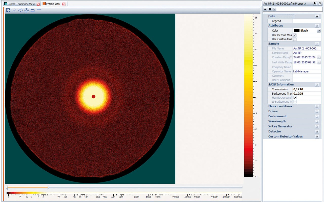
DIFFRAC.SAXS displays 1D and 2D SAXS data, as well as Nanography maps. Supported data formats are brml, raw, ascii (two-column xy and three-column xye with different units), gfrm and tiff.
Data pre-processing
DIFFRAC.SAXS allows extracting the pure scattering signal of the nanostructures straightaway.
In a first step the sample transmission is determined using glassy carbon as a reference or based on the primary beam intensity ratio. 2D data are subsequently sliced or integrated through optimized algorithms into 1D data for further evaluation.
DIFFRAC.SAXS can perform both the transmission calculation and the background correction automatically, without the need for user intervention, if the data measurement strategy is set up with DIFFRAC.WIZARD.
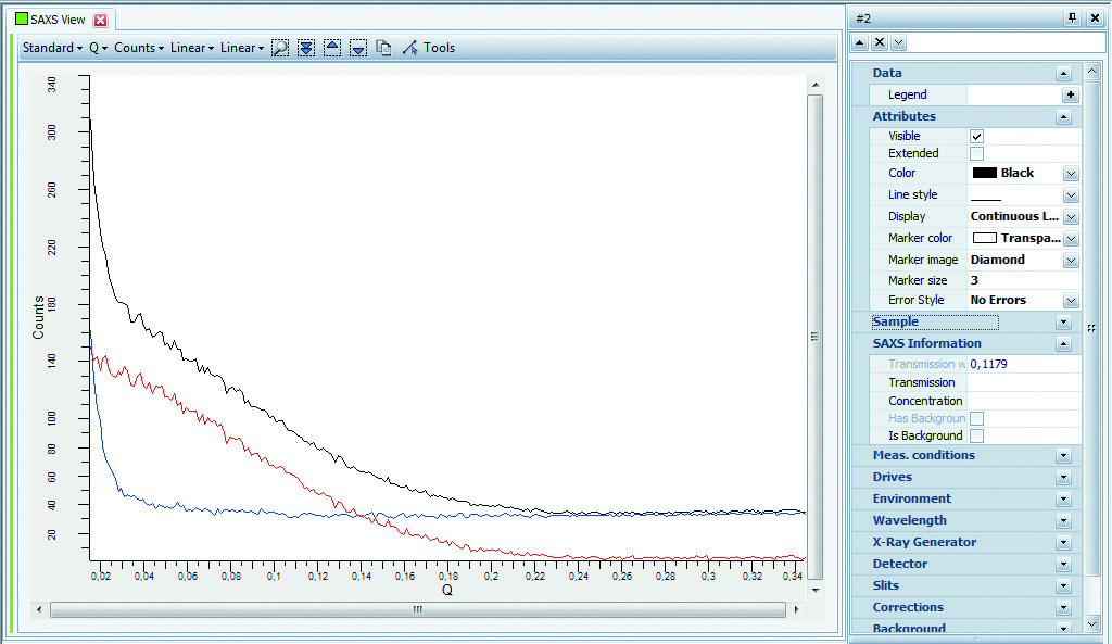
SAXS specific plots and evaluations
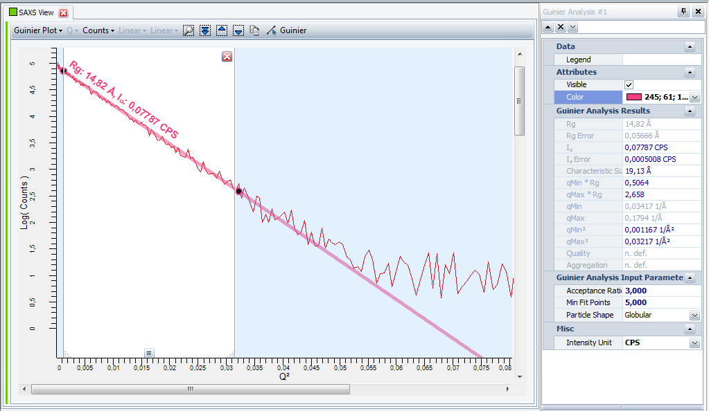
Traditionally, SAXS curves are displayed as plots with specific axis scaling that allow a quick graphical check of the data quality, and straightforward parameter evaluation such as the Guinier, Porod and Kratky plots. In DIFFRAC.SAXS all plot types can be readily selected by name. Evaluation of the related parameters, such the radius of gyration (Rg), the forward scattered intensity, the Porod scattering invariant, surface-to-volume ratio etc. is done fully automatically or interactively in a step-by-step procedure.
Model-based fitting
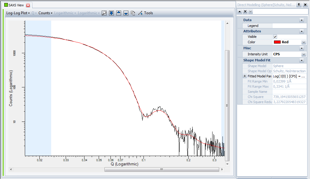
DIFFRAC.SAXS features a non-linear least-squares fitting of 1D SAXS data based on direct modeling of nanostructures in solution using geometrical shapes (sphere, cylinder, etc.) or by dedicated polymer models (chains, Gaussian star, etc.). It also takes into account polydispersity and concentration effects for complete sample characterization. The corresponding model parameters are determined by fitting the experimental SAXS curve.
Pair Distance Distribution Function
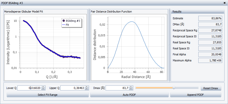
The Pair Distance Distribution Function (PDDF) p(r) is the distribution of the distance taken between any two points within the scattering particle. The PDDF is function of the size and shape of the particle and relates to the scattering curve by a sin Fourier transform.
DIFFRAC.SAXS Specifications
| Version | The current version of the software is DIFFRAC.SAXS V1.0. |
| Analytical methods | Interactive and automatic Guinier analysis providing radius of gyration, forward scattered intensity, etc. Interactive or automatic Porod analysis providing the Porod scattering invariant, surface-to-volume ratio, etc. Kratky analysis Molecular weight of proteins by scaling to absolute units Pair Distance Distribution Function (PDDF) Non-linear least-squares fitting of 1D SAXS data based on nanoparticle shape models, taking into account polydispersity and interaction effects |
Keep your DIFFRAC.SAXS up to date
Free Maintenance Update
The free DIFFRAC.SAXS Maintenance Update renews your SAXS version to the most recent release. Regardless of your SAXS license level, you can always download the latest Maintenance Update from www.brukersupport.com, free of charge!
Download process
- Register at Bruker Customer Support
- Click on the "Software" button
- Search for the DIFFRAC.SAXS Maintenance Update
- Download the update
Bugfixes
By keeping your DIFFRAC.SAXS up to date, you will benefit from all bugfixes made for the current but also all previously released versions, regardless of the license level. DIFFRAC.SAXS Maintenance Updates are cumulative and can therefore be applied to any previous version.
What are Upgrades?
DIFFRAC.SAXS Maintenance Updates do not come with new features. If you want to benefit from features introduced in new major releases you need to purchase the latest DIFFRAC.SAXS upgrade.