Research Highlight:
Kara Cerveny, Ph.D.
Reed College in Portland, Oregon
Light-Sheet Fluorescence Microscopy as a Tool to Study Development in the Visual System
This research highlight features Dr. Kara Cerveny, Associate Professor of Biology at Reed college. Dr. Cerveny discusses her experimental objectives using the light-sheet fluorescence microscope to study zebrafish eye development, including imaging the plane of cell division in real-time.

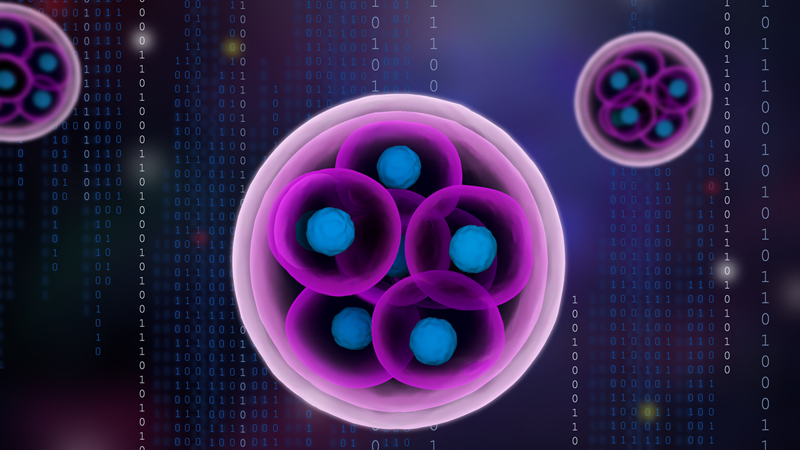
“The light-sheet is going to be amazing for us to be able to follow the plane of cell division in real time, actually watching dividing cells and seeing how the angle of cell division shifts over time, in response to various local cues. The light-sheet microscope being so gentle and fast is really nice for us, because with the confocal we have some preliminary data, but our time resolution was so limited. Because the light-sheet is super-fast, we can catch those divisions and really know if more proteins are being inherited on one side or the other, for instance, and really know precisely how the cell is dividing.”
Kara Cerveny, Ph.D., Associate Professor of Biology
Reed College in Portland, Oregon
ABOUT THE RESEARCHER:
Kara Cerveny, Ph.D., is an Associate Professor of Biology at Reed College in Portland, Oregon. She earned her B.S. in Biology and B.A. in Chemistry from Duke University and her Ph.D. in Biochemistry, Cellular, and Molecular Biology from Johns Hopkins School of Medicine.
FIELD OF STUDY:
Dr. Kara Cerveny’s research focuses on answering the question "how do cells transition from a proliferation state to a differentiation state?" using a light-sheet microscope to investigate the visual system, more specifically looking into how neuronal stem and progenitor cells behave in a growing zebrafish retina.