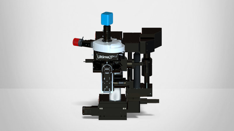
OptoVolt Module
Enabling Neural Millisecond Imaging
Bruker's OptoVolt™ Module for Ultima 2Pplus multiphoton microscopes delivers unprecedented kilohertz imaging rates to enable voltage imaging neural research. It provides a significant improvement over standard imaging techniques by capturing electrical dynamics greater than 1,000 frames per second and providing software-selectable multiplication modes.
These accelerated imaging speeds allow neuroscientists to capture millisecond dynamics of cell-to-cell neural communication, and with the emergence of fluorescent voltage indicators, it is even possible to measure neural activity with higher temporal resolution than fluorescent calcium indicators have allowed.

How the OptoVolt Module Enhances Ultima 2Pplus Performance
In this video: Jimmy Fong (Director, Products and Technology, Bruker Fluorescence Microscopy) walks through how the OptoVolt Module boosts imaging speed, expands experimental capabilities, and integrates seamlessly into existing Ultima 2Pplus systems.
You’ll learn how its scan‑multiplication optics enable true kilohertz‑rate voltage imaging and what that unlocks for high‑speed neural dynamics research. The overview also highlights how the module works, what it adds to your system, and the kinds of experiments it makes possible.
Contact us or continue reading to learn more.
Enhancements in Speed and Resolution
OptoVolt contains multiplication optics that re-route the resonant scanning beam for customized aspheric microlens arrays. It also leverages large detection optics for collecting scarce photons, in vivo and deep into tissue.
This module multiplies up the scan speed with the microlens array and the resulting multiplication factor is based on the number of microlenses scanned across. It integrates seamlessly with the Ultima 2Pplus, and is selectable as a scanning mode for ease-of-use. This gives researchers fast and flexible imaging for a range of novel experiments, including single-cell electrophysiology and wide-field calcium imaging. Researchers can build upon their existing microscope system as their research develops.


Traditional Resonant Raster vs. OptoVolt Scan
Without the OptoVolt Module, Ultima 2Pplus scans one line per sweep. This is repeated for as many lines as are wanted to create an image.
With OptoVolt's scan multiplier technology, instead of directly scanning this one line in the image, it scans through the microlens array. For every lens, it creates a multiplication factor.
As a result — as shown in this comparison — the OptoVolt module scan (green) can scan 16 lines during the same time it takes to sweep one line using the traditional resonant raster (red).
Advancements in Functional Mapping of Neural Circuits
The module leverages advanced voltage indicators, such as FORCE1s, and retains the depth and resolution of traditional two-photon imaging while enabling flexible adjustments between speed and signal intensity. A single resonant frame can be captured at 30 frames per second (fps), while a smaller strip can be imaged at several hundreds of fps. OptoVolt in 8x mode provides a larger field of view at a 565 fps rate, and can address potentially hundreds of cells, while 16x mode offers significant speed at 1130 fps.
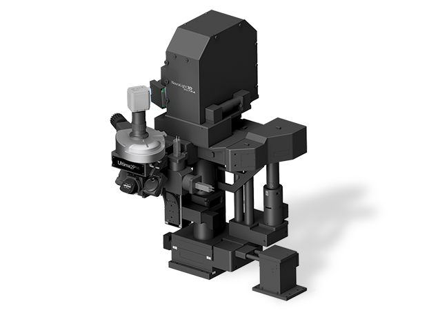
OptoVolt can also be integrated with the NeuraLight 3D Ultra SLM for simultaneous 3D optogenetic manipulation and voltage imaging, facilitating comprehensive all-optical functional mapping of neural circuits. This capability allows researchers to visualize action potentials and sub-threshold membrane potentials across large neural networks, bridging the gap between single-cell electrophysiology and wide-field calcium imaging.
This innovative module retains the depth and resolution of traditional two-photon imaging while enabling flexible adjustments between speed and signal intensity. Its design provides fast, efficient software switching between traditional Galvo, resonant, and OptoVolt modes for flexible control over any experiment.
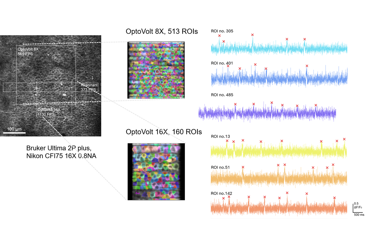
A full resonant frame, or a reduced resonant window (left), compared to example fields of view from mouse hippocampal CA1 cells using the voltage indicator FORCE1s and OptoVolt in 8x and 16x mode. The corresponding number of identified ROIs as imaged from CA1 (middle). Sample traces from the 8x ROIs (right).
Data courtesy of Qiyuan Liang of Michael Häusser’s lab (Hong Kong University), and François St-Pierre (Baylor College of Medicine).
Explore Emerging Applications
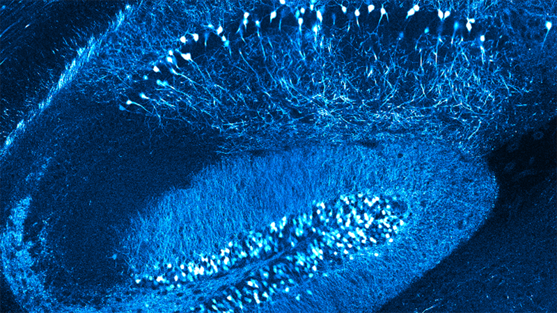
OptoVolt Reveals Precise Spatiotemporal Relationships of Hippocampal CA1 Neurons
This high speed video captured using the OptoVolt Module shows the propagation of millisecond voltage events across the brain area. Since image data is produced at every timepoint, researchers can dissect the spatiotemporal features of the voltage dynamics such as localized or coordinated activity.
Frequently Asked Questions
High speed data collection for fast dynamics can performed using point or line scan techniques, which are software features currently found in Prairie View software for the Ultima 2Pplus. These techniques can generate intensity time profiles for discrete excitation spots sequentially or along a single line with high temporal resolution.
In comparison, with the high speed scanning capability of the OptoVolt Module in both X and Y axes, an actual image is created across the FOV, which can reveal spatiotemporal dynamics across a larger area.
Bruker’s Ultima 2Pplus has entered into an exclusive agreement to market and commercialize the technology.
While the Ultima platform has been used successfully for fast voltage imaging, the dynamics measured in the standard Ultima 2Pplus with OptoVolt can becaptured with a much larger FOV, while maintaining the required imaging frame rate.
Yes, OptoVolt is compatible with your existing Ultima 2Pplus microscope. Since OptoVolt is based on the resonant scanner technology, an upgrade would require an upgrade to the Bruker resonant scanner module, if not already present. Please contact our representatives to inquire about the upgrade path toward the OptoVolt Module.
Yes, OptoVolt can be installed onto an existing Ultima 2Pplus microscope in the field by our field engineers without need to ship components back to the factory.
To maximize the effectiveness of the OptoVolt Module, the Ultima 2Pplus electronics platform would need to be updated to the Core Edition electronics if not already present. As with typical multiphoton imaging, we do recommend a dark room, with a vibration-isolated optical table to optimize the imaging quality.
OptoVolt can be combined with most modules on the Ultima 2Pplus, most notably the Neuralight 3D Ultra for combined 3D holographic optogenetic stimulation. Please consult your Bruker representative for possible unique combinations with other modules.
Please download the datasheet or contact your Bruker representative for more information, or to learn about opportunities to see the Ultima 2Pplus and OptoVolt in action.
Select Specifications
| Compatibility | Ultima 2Pplus with Resonant Scanner |
| Detection System | Bruker rtCore with ioCore electronics |
| Software | Prairie View version 6.0 or later |
| Multiplication Modes | Software selectable: x16: 1,100 fps; x8: 550 fps |
| Wavelength | Optimized 920nm, 700 to 1,100 nm compatible |
| Transmission | Minimum 70% |
| FOV | Objective dependent, size may vary depending on lens manufacturer Accessible Reference FOV: Scan Multiplied FOV area: |
| IP License | Boston University, Exclusive License: Scan Multiplier Technology for High-Throughput Scanning |
| DOWNLOAD THE DATASHEET FOR MORE INFORMATION | |