Differentiation of Protein Secondary Structure in Clear and Opaque Human Lenses: AFM–IR Studies
C. Paluszkiewicz, N. Piergies, P. Chaniecki, M. Rekas, J. Miszczyk, and W. M. Kwiatek
Key points
- AFM-IR was used to study secondary structure changes associated with cataract development
- The protein secondary changes from a β–turn/β–sheet parallel to an anti-parallel β–sheet as the lens is degraded
- Degraded regions were found to be stiffer than clear regions
Abstract
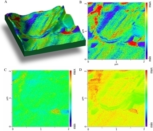
Here, we present the first approach to human lenses investigations with and without cataract development changes in nanoscale resolution using AFM-IR spectroscopy. We proved that the application of this technique allowed us to better understand of structural changes connected with advancing disease process in studied lenses.
The obtained results show the impact of the disease development on the secondary structure of proteins in these human tissues. The domination of the β–turn protein secondary structure is observed in the clear (non-affected by cataract) lens. While, in the case of the opaque (cataractous) samples the different degree of the degradation due to development of cataract, was recognized. Briefly, this process is associated with the protein secondary changes from β–turn/β–sheet parallel for less altered part of the lens to stable anti-parallel β–sheet for the more degraded part. Interestingly, the AFM-IR technique provided estimation of the protein secondary structure without the need for using deconvolution procedure.
Life Sciences

Nanoscale Infrared Spectroscopy
Measure spatially varying physical and chemical properties with nanoscale spatial resolution