Guide to Raman Microscopy
What is Raman Microscopy?
Raman microscopy (µ-Raman) combines the chemical analysis technique, Raman spectroscopy, with a traditional light microscope. This combination provides a very streamlined, “point and shoot” approach to Raman spectroscopy of very tiny objects. Users can directly select regions of interest on a sample for chemical analysis by visualizing them via microscope.
How Does a Raman Microscope Work?
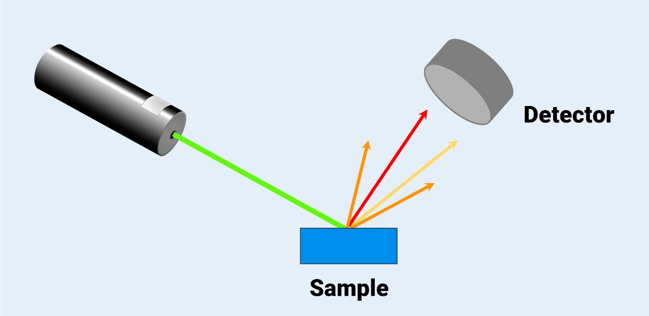
Raman microscopy relies on the same technique as Raman spectroscopy to identify the chemical compounds in a sample. First, a laser (typically a green 532 nm laser) illuminates the sample, interacts with it, and the laser light scatters off the sample’s surface. The laser light can be scattered in two ways: Rayleigh scattering or Raman scattering.
The Raman scattered light is the type that is useful for chemical analysis. This type of scattering is inelastic, which means the Raman scattered light has a different frequency, wavelength, and color then the original beam of light.
During a Raman experiment, the Raman scattered light is detected to create the Raman spectrum. This spectrum is unique to the chemical compounds present in the sample, so it acts as “chemical fingerprint” which can be used to identify, quantify, and characterize all the different chemical compounds present in the sample.
Using Raman, we can analyze anything as long as the laser light can reach it. This means Raman microscopy can analyze any sample that can be placed on the sample stage, with no sample preparation at all. It can even be used to analyze samples that are surrounded by glass, inside transparent packaging, or in water.
Raman spectroscopy does have a couple limitations, which apply to Raman microscopy as well. While Raman can be used to analyze any type of sample, some chemical compounds just don’t have a strong enough Raman signal to get a high-quality spectrum, so they can’t easily be analyzed with Raman spectroscopy.
Also, fluorescence is a big problem in Raman spectroscopy. If a sample shows fluorescence, the emitted light is much stronger than the Raman effect, making the Raman peaks difficult to distinguish from the noise.
Inside the Raman microscope
Since most Raman experiments use a laser in the visible range, it’s quite easy to combine a Raman spectrometer with a microscope. Afterall, microscopes are already designed to work with visible light. The only consideration here is the type of microscope to use, as there are many different microscope designs. Most Raman microscopes use a confocal microscope design to maximize resolution and spectral quality.
So naturally, we’ll cover the confocal design on our website.
The first part of the microscope is the sample stage. Once the sample is placed on the stage, we can view it with the microscope. From there, we can select a region of interest for analysis.
Light from the laser is then directed to the sample and focused with the objective lens. The laser light interacts with the sample and scatters off it, and all the scattered light travels back through the objective lens.
This scattered light consists of both Raman and Rayleigh scattered light, but only the Raman light is useful for chemical analysis. So, the Rayleigh light is removed with a filter. The remaining Raman scattered light is focused with a confocal pinhole and directed to the spectrometer, which separates the Raman light by wavelength and detects it.
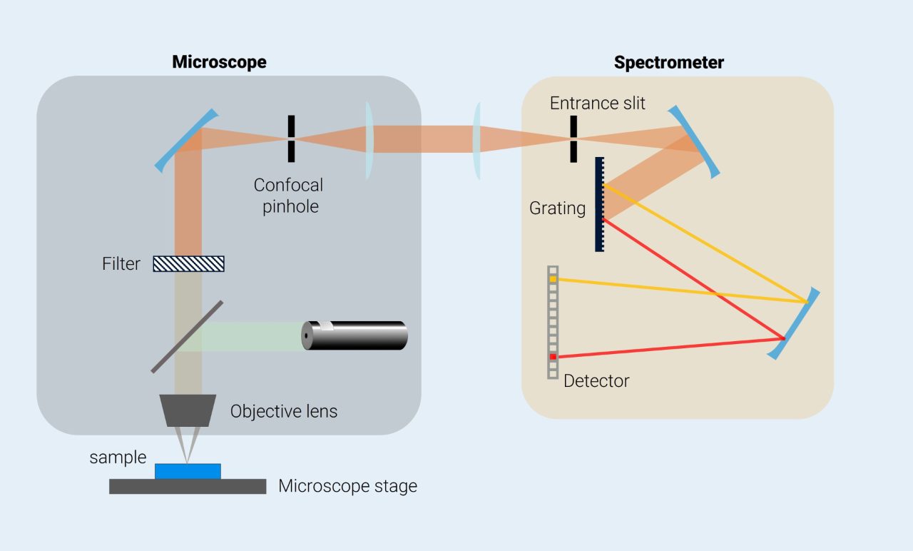
Once the Raman scattered light is detected, the Raman spectra is created. The Raman spectra for the region of interest contains a wealth of chemical information that can be used to identify and characterize the chemical compounds in that region of interest. Using computer software, these chemical compounds in the region of interest can even be identified automatically.
Lasers
Though the most common laser used in Raman Spectroscopy and Microscopy is a green 532 nm laser, that’s not the only option. Other commonly used lasers for Raman experiments include a 488 nm blue laser, 633 nm red laser, and 785 nm infrared laser. Different samples might need different lasers to maximize sensitivity and resolution, and to avoid fluorescence. That’s why many Raman microscopes have several lasers and being able to switch quickly and easily between them, is a must!
Objective lenses
Since the light in a Raman microscope usually travels back through the objective lens on its way to the detector, the objective lens has an important impact on the spatial resolution of the Raman microscope. In particular, the numerical aperture of the objective lens determines how much light can travel through the objective lens as well as the focal depth. The higher the numerical aperture of the objective lens, the higher the spatial resolution of the Raman microscope.
Gratings
The grating is the part of the spectrometer that separates the light by wavelength prior to detection. A Raman instrument usually houses multiple gratings which have different line densities. The line density of the grating corresponds to how much the light gets spread across the detector. How much the light spreads changes the spectral resolution, or how well we can see the individual peaks on the Raman spectra. Changing the spectral resolution can help us examine specific parts of the Raman spectrum in detail, or fine tune an experiment for different lasers.
About the confocal design
Earlier, we mentioned how most Raman microscopes are confocal microscopes, but what is a confocal microscope and why does it offer advantages in Raman microscopy?
When we use the laser to illuminate a spot on the sample, the laser doesn’t just interact with the surface. Rather, it can probe quite deep into the sample, depending on what the sample is made of. However, when we select a region for analysis, we only want the light that corresponds to the focal plane we’re investigating.
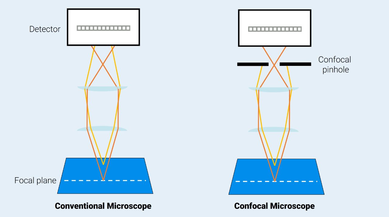
This is the advantage of the confocal microscope. This microscope design uses a pinhole to block out of focus light from reaching the detector. By removing the unfocused light, we can improve the spatial resolution, or how “sharp” the image produces by the microscope is. This is particularly useful in Raman imaging, where we need high spatial resolution to create detailed chemical images.
The confocal pinhole can also improve the quality of the Raman spectra itself by removing light that’s from fluorescence. While we can usually mitigate fluorescence by using a laser with a different wavelength, that’s not always a possibility. So, by removing the light from fluorescence, we can more easily get a clean Raman spectrum.
A final advantage of the confocal pinhole is that we can use it to move the focal plane. That is, we can selectively analyze the light corresponding to different depths in the sample so we can analyze a sample in three dimensions.
Types of confocal microscopes
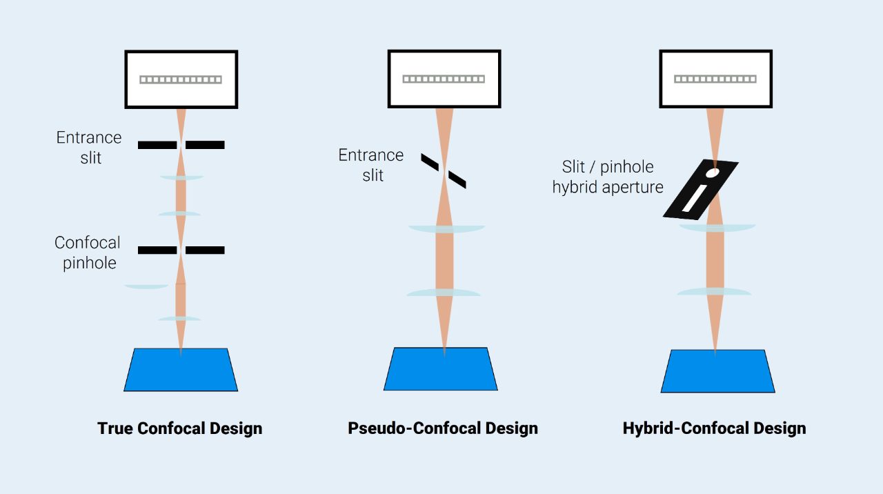
There are some tradeoffs with a confocal microscope, so there are actually a few ways of implementing this design into a Raman Microscope: true confocal design, pseudo-confocal design, and a hybrid-confocal design.
The true confocal design works exactly as described above: a confocal pinhole removes out of focus light before it’s sent to the spectrometer, which improves the spatial resolution.
However, as the light enters the spectrometer, it also passes through a slit which further eliminates light. This entrance slit controls the spectral resolution, or how well we can see the individual peaks on the Raman spectra. The confocal pinhole and the entrance slit must be aligned so the microscope has good performance. However, this is difficult to maintain, which decreases the throughput of the microscope.
The pseudo-confocal design gets around this by removing the confocal pinhole. This design simulates the effect of the confocal pinhole by placing the entrance slit and detector orthogonally to each other to help remove that out of focus light. By simplifying the design of the microscope, the throughput is increased at the expense of the spatial resolution.
Both designs offer advantages, which is why the hybrid-confocal design combines these benefits to give access to true confocal microscopy or high throughput. It does this by using a hybrid array of pinholes and slits that can act as the entrance slit or the confocal pinhole.
Raman microscopy applications
Raman microscopy has two huge benefits: the convenience of a “point and shoot” approach to chemical analysis and the ability to analyze even the smallest objects (> 0.5 µm). These benefits mean Raman microscopy has uses in a wide variety of industries.For example, Raman microscopy can be used for quality control and failure analysis, where small defects can be selectively analyzed without damaging the sample.
Of course, Raman microscopes are also used for imaging to create chemical images. Chemical imaging with Raman allows us to distinguish chemical species that look the same when viewed with a microscope or view the distribution of chemicals in a sample.
Learn more about Raman imaging
The primary use of a Raman microscope isn’t to just create Raman spectra, it’s to create detailed chemical images. In these chemical images, each pixel contains a Raman spectrum. The various compounds in all those spectra are differentiated and assigned different colors. This allows the nature and structure of the sample to be visualized and analyzed precisely.

Frequently asked questions about Raman microscopy
What are advantages of Raman spectroscopy
Raman has several major advantages in comparison to other vibrational spectroscopy technique such as FTIR and NIR absorption. Opposed to absorption, Raman effect is the inelastic light scattering off a sample. As a result, Raman spectroscopy requires no or little sample preparation in measurement of solids, liquids, and gases. Not only directly, but also through transparent windows such as glass and plastic. Water has very low Raman signal and therefore Raman spectroscopy can easily detect compounds that dissolved in water without strong interference. That makes Raman spectroscopy very suitable for biological samples in native state.
How long does it take to acquire a Raman spectrum?
The exposure time depends on many factors, such as the expectation of spectral quality, laser power, and samples cross-section for Raman scatter. Typically, good-quality Raman spectra can be acquired within a few seconds.
Can Raman spectra be obtained from a mixture of materials?
The Raman spectrum contains information about all molecules that are measured. Therefore, Raman spectra obtained from mixture contain peaks from various molecules. If the spectra of components are known, quantitative information about the composition can be generated.
What else information can Raman detect, other than chemical structure?
Raman spectroscopy can provide, directly or indirectly, various information such as isotopes in molecules, allotropes, crystallinity, polymorphism, doping in crystal lattice, tension, pressure, and temperature.
Is Raman spectroscopy quantitative?
The intensity of a spectrum is linear to concentration. The relationship between peak intensity and concentration can be calibrated with known samples. In mixtures, Raman peaks provide quantitative information about the concentration of compounds at the same time.
What is the best laser wavelength for my application?
Unfortunately, the best laser wavelength for a specific application is also not always obvious. Many system variables must be considered to optimize the excitation wavelength in a Raman spectroscopic experiment. The scattering efficiency, influence of fluorescence, detector efficiency, as well as the availability of the cost-efficient and easy-to-use system, are the main aspects need to be considered. The resulting most used wavelength is 785 nm and/or 523 nm. The 532 nm is particularly suitable for inorganic materials, e.g. graphene and fullerenes.
What is the typical laser power for Raman measurements?
The laser power at the sample on a Raman microscope is typically from sub-mW level to a few tens of mW. The Raman intensity is directly proportional to the laser power. However, there is an increased risk of sample damage when using strong laser power. The laser power can be lowered to avoid sample damage but in doing this we need longer exposure time to acquire good-quality spectra.