Application Note: Using Scanning Electrochemical Atomic Force Microscopy to Image Redox Immunomarked Proteins on Viruses
In this application note, readers will explore how Mediator-tethered Atomic Force Microscopy–Scanning Electrochemical Microscopy (Mt/AFM-SECM) enables nanoscale electrochemical imaging of redox-labeled proteins on individual virus particles. By integrating the high spatial resolution of AFM with the chemical sensitivity of SECM, this advanced technique reveals molecular-level features that remain undetectable through topographical imaging alone.
Contents inlcude:
- The benefits of combining AFM and SECM for nanoscale protein mapping on virus particles
- How Mt/AFM-SECM uses redox-tagged antibodies and oscillating probes for in situ electrochemical detection
- Application examples in mapping coat proteins and VPg on plant viruses like LMV and PVA
Introduction
In Scanning Electrochemical Microscopy AFM (SECMAFM), a metal-coated AFM tip, insulated except its very apex, is used as an electrode to generate/detect chemical species in an electrolyte solution at a desired location (the principle is shown in Fig.1). The main interest of this technique is that it combines the standard capabilities of an AFM (such as generating a 3D-profile of the surface or applying a controlled force to the sample) to the possibility of electrochemically generating/ detecting species under a specific potential.
Thus the AFM probe can be used both as a force sensor and a microelectrode. Until recently, this technique had never been applied to characterize actual biological samples [1].
In the present application note, the tip has been brought into contact with redox immunomarked viral particles in order to electrochemically interrogate them. The aim was to achieve in situ mapping of specific viral proteins.
Viruses are natural nanomachines. In spite of their simple architecture, they can display complex activities and can infect both prokaryotic and eukaryotic cells. Over the last twenty years, they have also been used as nanovectors in nanomedicine [2], nano-vehicles for enzymatic catalysis [3] or scaffolds for complex constructs having structural and functional properties [4]. For nanotechnology applications, two types of viruses are often used: bacteriophages [5] and plant viruses [6], since they are harmless to humans. In this study, two filamentous plant viruses (Potyvirus gender), the lettuce mosaic virus (LMV) and the potato virus A (PVA), have been investigated by SECM-AFM. Until recently, the best resolution achieved in SECM was in the micrometer range [7], which is certainly enough to probe processes at a large scale (for instance on a living cell) but not to address nano-bio objects like viruses. This technical note describes a new technique called Mt/ (Mediator-tethered) AFM-SECM in which a microelectrode probe is used to electrochemically contact redox-labeled molecules attached onto an electrode surface [8]. This type of construct has been proven to enable detection of the dynamics of PEGs [9] (Polyethylene-glycols) or DNA chains [10]. Mt/AFM-SECM can be combined with redox immunomarking (the use of antibodies grafted with redox PEG chains) to specifically locate ~100 nm size antigens attached to a synthetic surface [11]. Measuring the activity in situ of redox-labeled, immobilized active biostructures requires both a nanometer range spatial resolution and the ability to measure very low electrochemical current (down to the tens of femto Ampere range).
Imaging of Redox Protein on Viruses
Potyviruses are rod-shaped particles of ~700 to 900 nm in length and ~10 to 15 nm in width made of helical winding identical coat proteins (CPs). For most virus imaging AFM experiments, mica is the substrate of choice, but in order to enable Mt/AFM-SECM measurements, the substrate has to be conductive. Hence LMV and PVA particles are deposited on a template-stripped gold surface. The viruses are then specifically immunomarked by primary antibodies and finally made "electrochemically visible" using secondary antibodies tagged with redox ferrocene (Fc)-PEG chains.
To perform Mt/AFM-SECM measurements, a homemade AFM-SECM tip is oscillated at its fundamental flexural frequency (~2-3 kHz), biased at Etip = +0.3 V/SCE and brought close to the surface which was biased at Esub = 0.0 V/SCE. The approach was stopped and raster imaging started when the cantilever oscillation was damped by 10%. Topography and tip current images were simultaneously recorded.
Fig. 2 shows a representative example of what can be obtained on CP-marked LMV particles. On the topography channel (a) and the corresponding longitudinal cross-sections (d: red traces), the capsids appear to be mostly homogeneous and featureless. By contrast, the tip current images (b) captured simultaneously and the corresponding cross-sections (d: blue traces) reveal a heterogeneous profile, in the sense that well-defined current spots are present along the virus capsids. This phenomenon is even more obvious on the 3d-rendering (c and e). These clearly defined spots are due to clusters of anti-CP/Fc-PEG IgG immunocomplexes bound to the virus capsid. The different intensities can be attributed to a variable number of Fc heads (according to the authors, up to 3 IgG-PEGFc molecules are able to bind the anti-CP antibody).
It is very interesting to note that the immunocomplex clusters cannot be detected in topography. The fact that antibody clusters are only detectable in the tip current images shows all the interest of Mt/AFM-SECM as a technique of high potential for mapping immunomarked proteins on virus particles. Moreover, those results indicate that Mt/AFM-SECM can reveal the way redox functionalization is distributed not only among the viruses but also over individual viruses, a valuable feature for viral nanotechnology applications.
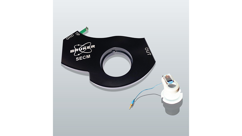
For such a project, the JPK Nanowizard systems are particularly suitable, especially with respect to the open access to the cell. Starting from the standard configuration (using the regular glass tip holder), a wire was added and connected to a home-designed bipotentiostat. The new setup was also softwareimplemented. What is also unique to the JPK configuration is the presence of a real reference electrode which is the key requirements for all electrochemical AFM measurements.
Highest Achievable Resolution
Apart from the CP, another protein known as VPg (Viral Protein linked to the Genome) is present in the virus capsid. This protein is covalently linked to the 5'-end of the virus RNA and is located at the corresponding end of the particle. In literature, it is postulated that a part of VPg protrudes from the particle surface [12]. In order to check this assumption, VPgs were specifically redoximmunomarked on LMV particles, and the same technique was used to image them.
Fig. 3 shows an inset of an image of two LMV particles (topography in a and tip current in b). The 3d-rendering (c) clearly shows a protrusion of about 30 nm in diameter that is in perfect compliance with the expected size of the immunocomplex (see schematic in e). The tip current profile (blue line in d) specifically shows the presence of the immunocomplex cluster formed at the VPg-exposing extremity of the virus.
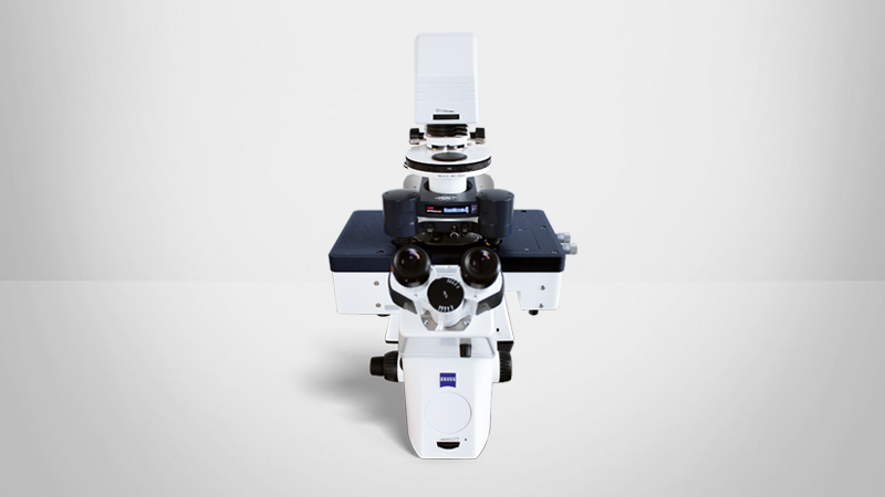
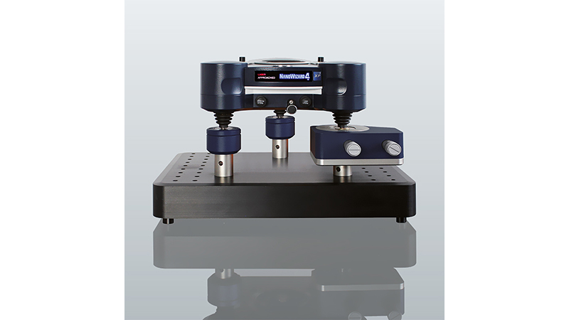
In figure 4 is shown the recently released Nanowizard 4 AFM, on an inverted optical microscope. Such setup can be used for AFM-SECM experiments.
Conclusion and Perspectives
Mt/AFM-SECM has been used to image in situ proteins on individual virus particles with a current detection sensitivity of ~10 fA and a spatial resolution of ~10 nm, such that single-protein molecules can be detected. This type of approach can very well be extended to the investigation of bioactive nanometric devices mimicking the high complexity of plasma membranes or even living cells. As an example, viruses may be used as scaffolds to specifically target and bind redox enzymes. In that respect, Nanowizard AFMs operated in scanning electrochemical mode are perfect tools for viral nanotechnology in the sense that they uniquely allow the functional characterization of modified viruses.
References
- "Combined Atomic Force Microscopy Scanning Electrochemical Microscopy. " C. Demaille, A. Anne, In Nanoelectrochemistry. M.V. Mirkin, S. Amemiya. Eds, Taylor and Francis Group, p749-788.(2015).
- "Nanoparticles as Platforms for Next-Generation Therapeutics and Imaging Devices" N.F. Steinmetz, Nanomedicine, 6 634-641 (2010).
- "Virus Scaffolds as Enzyme Nano-Carriers" D. Cardinale, N. Carette T. Michon, Trends Biotechnol., 30 369-376 (2012).
- "Reengineering Viruses and Virus-like Particles through Chemical Functionalization Strategies" M.T. Smith, A.K. Hawes and B.C. Bundy, Curr. Opin. Biol., 24 1089-1093 (2013).
- "Programmable Assembly of Nanoarchitectures Using Genetically Engineered Viruses" Y. Huang, C.Y. Chiang, S.K. Lee, E. Hu, J. De Yoreo and A.M. Beicher, Nano Lett., 5 1429-1434 (2005).
- "Utilization of Plant Viruses in Bionanotechnology" N.F. Steinmetz and D.J. Evans, Org. Biomol. Chem., 5 2891-2902 (2007).
- "Scanning Electrochemical Microscopy Imaging" F.R.F. Fan, Scanning Electrochemical Microscopy, 2nd Ed., (2012).
- "Electrochemical Atomic Force Microscopy Imaging of Redox-Immunomarked Proteins on Native Potyviruses: From Subparticle to Single-Protein Resolution" L. Nault, C. Taofifenua, A. Anne, A. Chovin, C. Demaille, J. Besong-Ndika, D. Cardinale, N. Carette, T. Michon and J. Walter, ACS Nano, 9 4911-4924 (2015).
- "Probing the Structure and Dynamics of End-Grafted Flexible Polymer Chain Layers by Combined Atomic Force-Electrochemical Microscopy, Cyclic Voltametry within Nanometer-Thick Macromolecular Polyethylene-glycol Layers" J. Abbou, A. Anne and C. Demaille, J. Am. Chem. Soc., 126 10095-10108 (2004).
- "Exploring the Motional Dynamics of End-Grafted DNA Oligonucleotides by in-situ Eletrochemical Atomic Force Microscopy" K. Wang, C. Goyer, A. Anne and C. Demaille, J. Phys. Chem.B, 111 6051-6058 (2007).
- "High resolution Mapping of Redox-immunomarked Proteins Using Electrochemical Atomic Force Microscopy in Molecule Touching Mode" A. Anne, A. Chovin, C. Demaille and M. Lafouresse, Anal. Chem. 83 7924-2932 (2011).
- "An Unusual Structure at One End of Potato Potyvirus Particles" L. Torrance, I.A. Andreev, R. GabrenaiteVerhovskaya, G. Cowan, K. Mäkinen, M.E. Taliansky, J. Mol. Biol. 357 1-8 (2006).


