Fragment Screening by Ligand Observed NMR
In addition to Surface Plasmon Resonance (SPR) and X-ray crystallography, Ligand observed NMR is one of the most popular techniques used in Fragment Based Drug Discovery (FBDD). In recent years methodological and technical advancements have enabled NMR-based fragment screening to be performed in full automation and with significantly reduced consumption of fragments and unlabeled target protein. Ligand observed NMR can not only be used as a primary screening tool but also to confirm hits from other screening methods such as Thermal Shift, which results in high quality hits. Furthermore, all experiments can be performed as competition experiments in which a known binder is displaced and the Kd is determined by titration.
Here we present practical aspects on how to setup ligand observed NMR screening experiments in an automated fashion for the three basic experiments: Saturation Transfer Difference (STD), Water-Ligand Observation with Gradient SpectroscopY (waterLOGSY) and relaxation based methods (T1r and T2).
Ligand Observed NMR Screening Experiments:
- Large molecules > 20 kDa exhibit significantly different NMR properties in terms of relaxation (T1 and T2) and Nuclear Overhauser Effect (NOE) than fragment like small molecules.
- It is common to all ligand observed screening experiments, that ligands which bind to the target can be distinguished from non-binders due to interaction with a macromolecule which in turn changes the fragment’s observable NMR parameters.
- Hits detected with NOE-based methods can be confirmed by using a relaxation based method.
- During the residence time on the protein and due to chemical exchange when binding, small molecules adopt relaxation properties of the target molecule. This can be used to distinguish binders from non binders.

T2/T1r – Relaxation based methods
T2 and T1r relaxation times of large biomolecules are much shorter than those of fragments. Usually two spectra are measured and compared, one with 10 ms relaxation delay and one with 200 ms relaxation delay. Fragments that bind can be identified from a comparison of the spectra:
waterLOGSY
In WLOGSY experiments polarization is transferred from water molecules to small molecules by NOE. Binding ligands interact with water that is bound to the protein, while non-binders only “see” the solvent water. The NOE effect, however, from bound water has the opposite sign than the one from free water. As a result, the signal from binding fragments is opposite to signals from non-binders.
Saturation Transfer Difference (STD)
In STD experiments, two spectra are recorded, (1) a spectrum with selective radio frequency (RF) irradiation on the biomolecular target, and (2) a spectrum with off-resonance irradiation.
Saturation of the target due to RF irradiation is transferred to binding ligands via NOE, resulting in a reduced intensity in the on-resonance spectrum. In a difference spectrum of (1) and (2) only signals from binders can be observed.


Key Advantages of NMR Screening
- NMR techniques do not require protein specific setup or knowledge about the protein’s function and can be applied to targets for which no bio-assay is available,
- NMR techniques in solution are not compromised by crystal contacts or target immobilization,
- weak binding from low uM to mM can be detected
- QC of fragments, easy identification of degradation in spectra
Experimental Parameters
Current TopSpin releases provide dedicated parameter sets for automated ligand-observed fragment screening workflows. Available experiments include waterLOGSY, STD, T1ρ, T2 (CPMG), and 19F T2 screening, enabling robust detection and validation of protein-ligand interactions across a wide range of targets.
Available screening parameter sets include:
- SCREEN_WLOGSY: WaterLOGSY-based screening experiment
- SCREEN_STD: Saturation Transfer Difference (STD) screening experiment
- SCREEN_T1R: T1ρ-based relaxation screening experiment
- SCREEN_T2: CPMG-based T2 relaxation screening experiment
- SCREEN_19F_T2: CPMG-based 19F screening experiment for fluorinated compound libraries
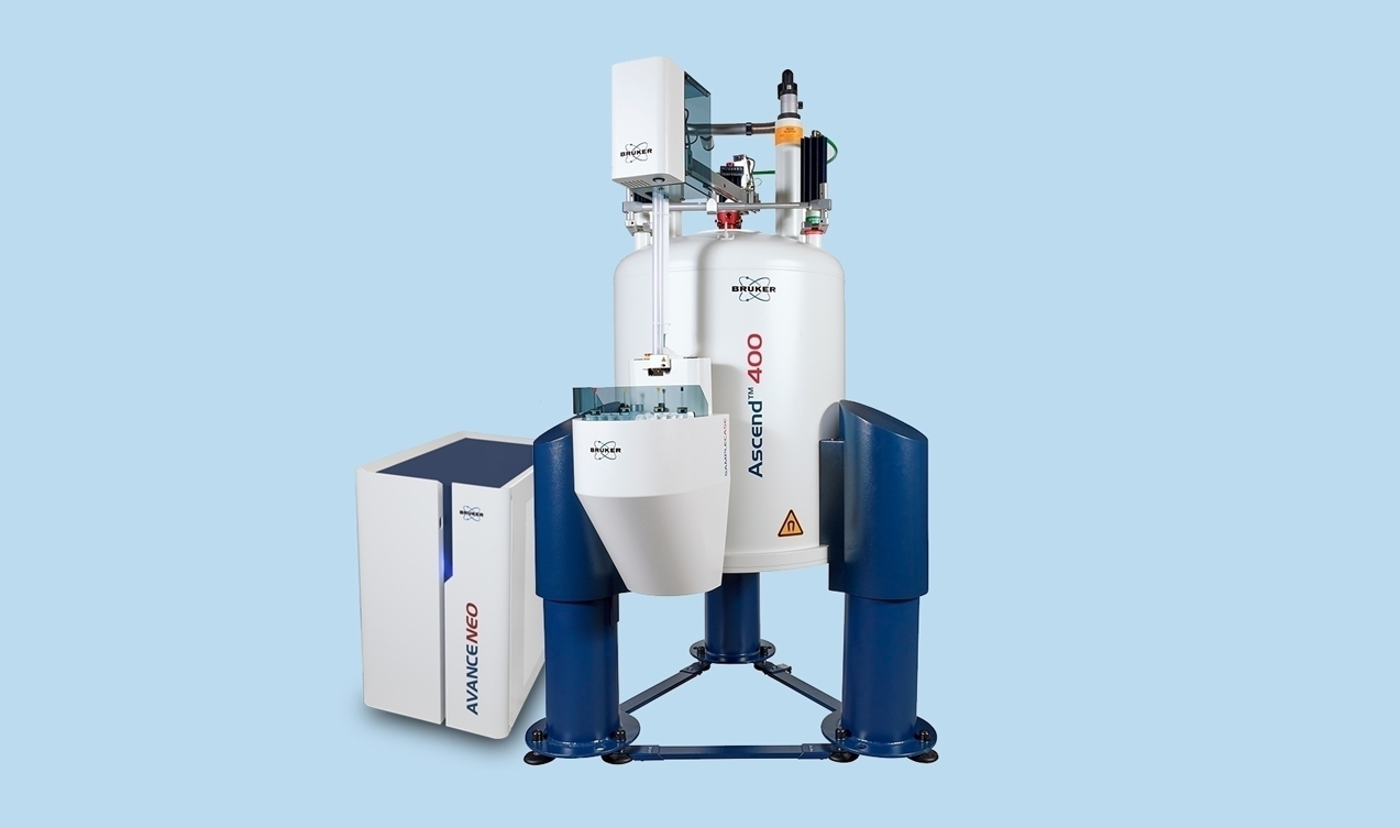
Ligand-observed screening experiments can be performed on standard high-resolution NMR systems equipped with automated sample handling, enabling efficient screening of large fragment collections and automated hit validation workflows.
- The diagram illustrates recommended protein concentration ranges for the different screening methods.
- Optimal protein concentration and ligand-to-protein ratios depend strongly on target molecular weight and binding affinity.
- Larger proteins generally provide stronger contrast between binders and non-binders, allowing higher ligand-to-protein ratios.
- For sample-limited targets, ligand-to-protein ratios of up to approximately 200:1 may be employed.
- The use of fragment cocktails significantly increases screening throughput. Typical cocktail sizes are fewer than 10 compounds for 1H screening and fewer than 30 compounds for 19F screening.
- Automated acquisition and processing routines facilitate standardized data collection and rapid identification of binding fragments.
Conclusion
Ligand observed NMR is an automated fragment screening technique that yields high quality hits in primary and secondary screens. Nowadays, parameter sets with optimized measurement conditions are available which significantly facilitates the setup of NMR-based screening experiments, even for novice users. In addition, a key advantage is the QC of fragments from 1D 1H spectra which enables the identification of compound degradation and aggregation, avoiding false positives. This is especially useful for confirmation of hits from primary screens performed by using SPR or Thermal Shift.
Further reading:
A.D. Gossert and W. Jahnke, Prog. NMR Spect. 2016
Authors:
Stefan Jehle1, Helena Kovacs1, Till Kuehn1, and Alvar Gossert2
1 Bruker BioSpin AG, Industriestrasse 26, 8117 Faellanden, Switzerland, 2Novartis Institutes for BioMedical Research, Basel, Switzerland.