Imaging Hippocampal Microcircuitry Using Glass Microperiscopes
Learn how leading researchers use 2-photon technology to see deeper connections between hippocampal microcircuit activity and behavior.
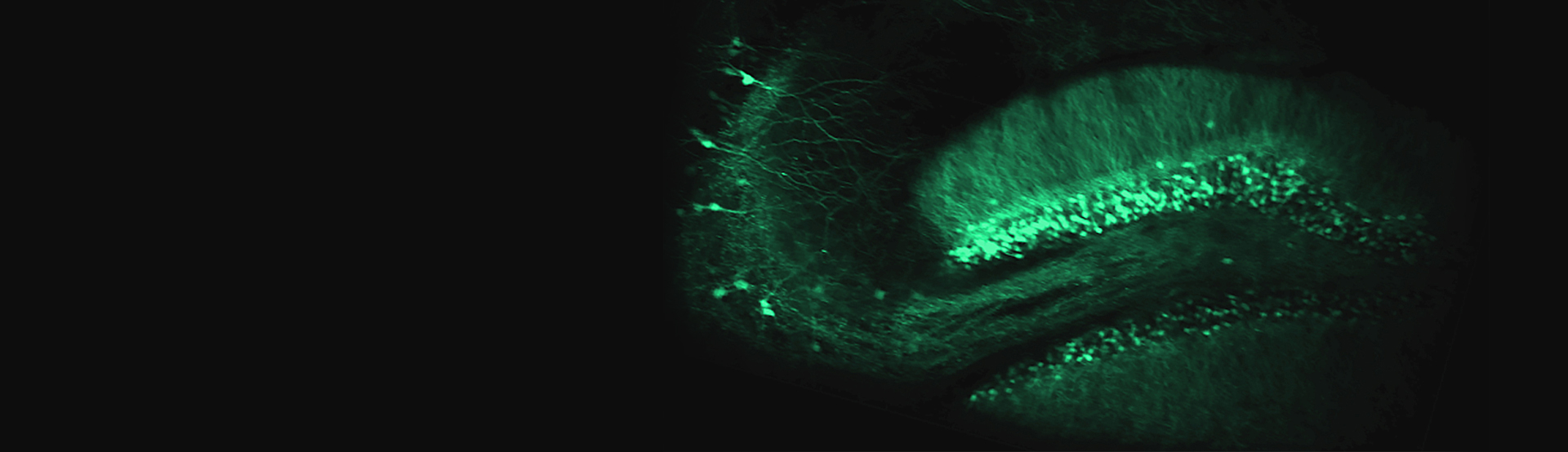
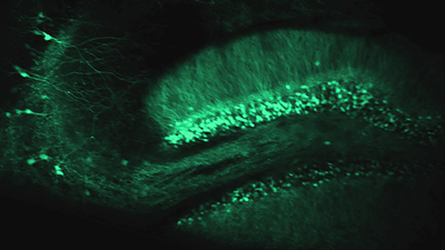
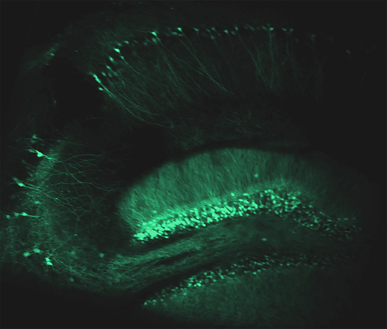
During this webinar, guest presenter Dr. Michael Goard (U.C. Santa Barbara) demonstrates a new approach for functional imaging of the transverse plane of the murine hippocampus
Viewers will gain direct insight into how Dr. Goard's lab uses this approach to measure dendritic morphology and functional activity during behavior, including descriptions of experimental procedures, examples of data collected, and a discussion of the advantages, limitations, and potential future applications of this approach.
Overview
Dr. Goard's research is a great example of the advantages of using two-photon technology for advanced neuroscience research and its potential uses as a foundation for novel research methods.
Watch the on-demand recording or download the webinar recap to find out more.
Presenter's Abstract
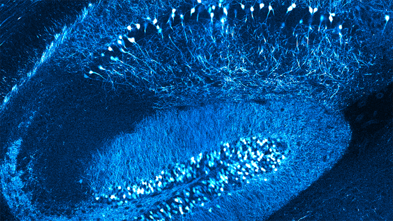
The hippocampus consists of repeated motifs of a canonical neuronal microcircuit first described by neuroanatomists over a century ago. This microcircuit contains distinct subfields (CA1-3, DG) with stereotyped connectivity that are critical for memory formation. However, optical imaging of the canonical circuit has not been possible in intact animals. To address this, our lab developed an approach for two-photon imaging of the transverse hippocampal plane in awake mice via implanted glass microperiscopes. This approach allows us to track dendritic morphology over repeated sessions and measure functional responses (e.g., place fields) using genetically encoded calcium indicators. In this talk, I will describe the procedure, imaging results, and discuss general considerations for the use of glass microprisms for deep brain imaging.
Featured Products and Technology
Speaker(s)
Michael Goard, Ph.D., Associate Professor, Department of Molecular, Cellular and Developmental Biology, Department of Psychological & Brain Sciences
Michael Goard received his B.A. in Psychology from Reed College and his Ph.D. in Neuroscience from UC Berkeley, where he completed his dissertation in the lab of Prof Yang Dan. As a postdoctoral fellow with Prof Mriganka Sur at MIT, he developed approaches for imaging the activity of large populations of neurons in behaving mice. He joined the faculty of UC Santa Barbara in 2016, where his lab investigates the neural circuitry underlying our perceptual, spatial, and cognitive abilities.