European BioAFM User Meeting [2022]
Discover leading-edge BioAFM research and the latest advances in BioAFM instrumentation.
An impressive panel of researchers share insights into their work using Bruker BioAFM technology.
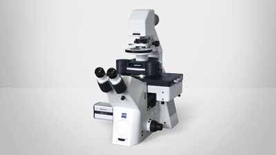
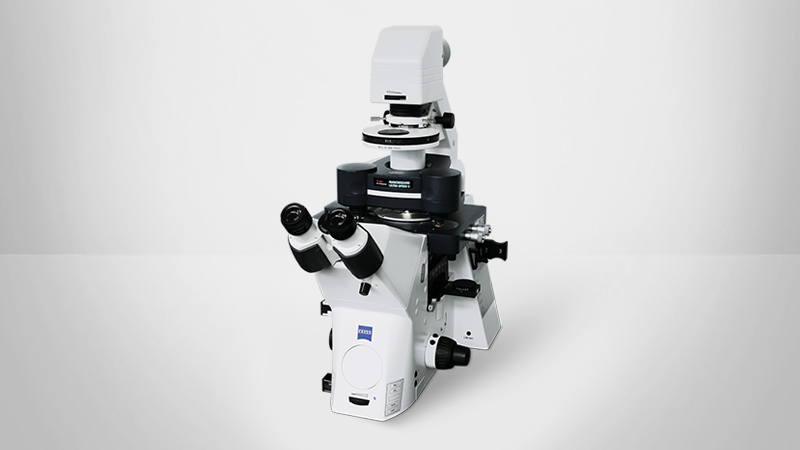
Featuring real-time demonstrations of state-of-the-art BioAFM instruments—including the newly launched NanoWizard V BioScience AFM—recorded LIVE from our laboratories in Berlin.
Find out more about the technology featured in this event or our state-of-the-art BioAFM technology:
Agenda
| Time (CET) | Topic | Speaker(s) |
|---|---|---|
| 14:00 | Welcome & Introduction | Peter Dewolf, Ph.D., Worldwide Application Director, Bruker Nano Surfaces and Metrology Thomas Henze, Ph.D., Head of Applications BioAFM, Berlin, Germany |
| 14:05 | Latest BioAFM Developments | Thomas Henze, Ph.D., Head of Applications BioAFM, Berlin, Germany |
Session 1: Mechanics, Large Samples, and Tissues
| Time (CET) | Topic | Speaker(s) | Abstract |
|---|---|---|---|
| 14:20 | Single Cell Compression Using AFM | Katarzyna Pogoda, Ph.D., Institute of Nuclear Physics, Polish Academy of Sciences, Poland | Read More |
| 14:45 | AFM and Fluorescence Microscopy on Human Osteoarthritic Cartilage: Correlation of Chondrocyte Organization, Nanomechanics and Collagen Structure Across Length Scales | Mathäus Tschaikowsky, Institute of Physical Chemistry, University of Freiburg, Germany | Read More |
| 15:10 | Live Lab Demo: NanoWizard V | Tanja Neumann, Ph.D., Bruker BioAFM, Berlin, Germany | |
| 15:30 | Coffee Break |
Session 2: High-Resolution & Fast Imaging, Super-Resolution Microscopy
| Time (CET) | Topic | Speaker(s) | Abstract |
|---|---|---|---|
| 15:35 | Phase Separation in the Outer Membrane of Escherichia coli | Georgina Benn, Ph.D., Silhavy lab, Princeton University, USA | Read More |
| 16:00 | AFM-STED Correlative Microscopy | Claudio Canale, Ph.D., Department of Physics, University of Genoa, Italy | Read More |
| 16:25 | Live Lab Demo: NanoWizard ULTRA Speed 2 | Andre Körnig, Ph.D., Bruker BioAFM, Berlin, Germany | |
| 16:50 | Q&A and Closing | Peter Dewolf, Ph.D., Worldwide Application Director, Bruker Nano Surfaces and Metrology Thomas Henze, Ph.D., Head of Applications BioAFM, Berlin, Germany |
Featured Products and Technology
Speakers and Abstracts
With introduction, Q&A, and conclusion led by Peter Dewolf, Ph.D., Worldwide Application Director, Bruker Nano Surfaces & Metrology, and Thomas Henze, Ph.D., Head of Applications BioAFM
Single Cell Compression using AFM
Katarzyna Pogoda, Ph.D., Institute of Nuclear Physics, Polish Academy of Sciences, Poland
ABSTRACT: During the talk, I will discuss how to perform uniaxial compression of single cells via Atomic Force Microscopy and how to obtain stress-strain curves to describe compression-stiffening behaviour in single cells.ABOUT THE PRESENTER: Dr. Katarzyna Pogoda has an M.Sc in Biophysics and holds a Ph.D. in Physics. She was a Fulbright Fellow and a Postdoc at the University of Pennsylvania, USA. Currently, she is employed as an Assistant Professor at the Institute of Nuclear Physics PAN, Poland. Dr. Pogoda has been working with Atomic Force Microscopy since 2010. In her work, she has been exploring the rheological properties of live cells in the context of their cancerous transformation.
AFM and Fluorescence Microscopy on Human Osteoarthritic Cartilage: Correlation of Chondrocyte Organization, Nanomechanics and Collagen Structure across Length Scales
Mathäus Tschaikowsky, Institute of Physical Chemistry, Albert-Ludwigs-University of Freiburg, Germany
ABSTRACT: While it is well established that many tissues, such as cartilage, consist of hierarchical structures, we still barely understand how different structural length scales determine the properties of these tissues. One reason for this is that, currently, nanoscale information can only be obtained very locally and often cannot be related to larger scales. This limits our ability to gain insights into the understanding of diseases related to biomechanics, such as osteoarthritis. Here, we demonstrate how a hybrid fluorescence-AFM setup provides both mechanical and topographic information of osteoarthritic cartilage with nanoscale resolution over a range of several millimeters.ABOUT THE PRESENTER: Mathäus Tschaikowsky is a doctoral researcher and part of Dr. Bizan Balzer’s team in the lab of Prof. Dr. Thorsten Hugel at the Institute of Physical Chemistry in Freiburg. In cooperation with G.E.R.N. lab (University Medical Center Freiburg), he is studying the nanomechanical and morphological changes in human articular cartilage at the early stages of osteoarthritis. Using Atomic Force Microscopy in combination with fluorescence microscopy, he could study the pathology of the tissue at the nanometer scale.
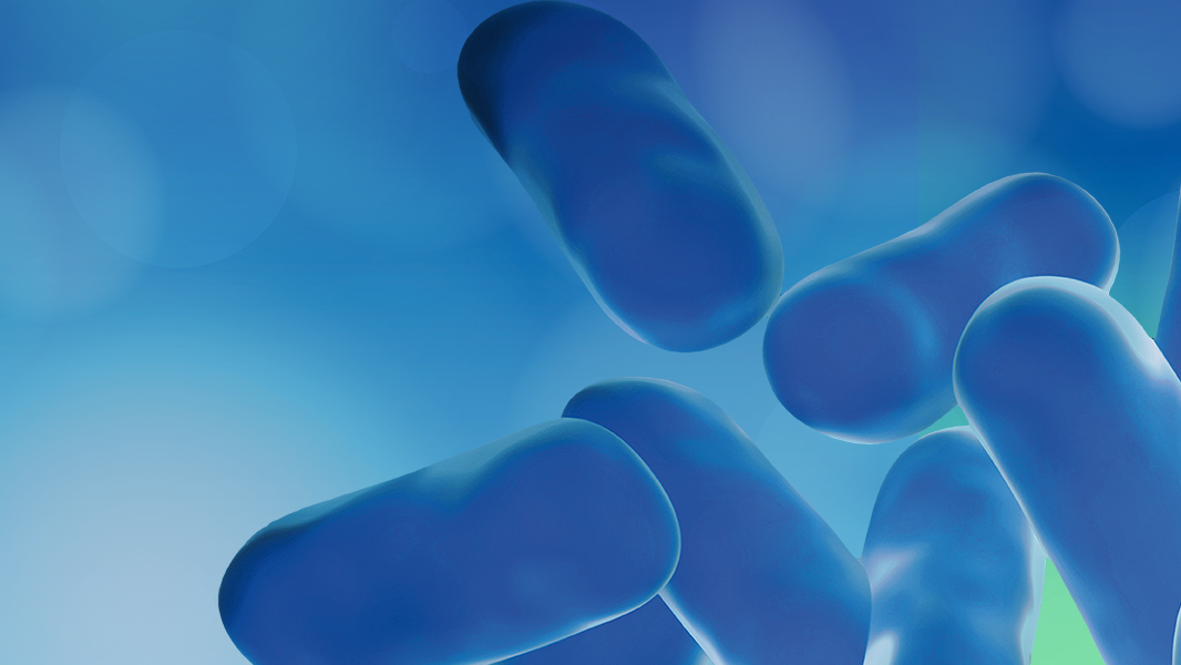
Phase Separation in the Outer Membrane of Escherichia coli
Georgina Benn, Ph.D., Silhavy Lab, Princeton University, USA
ABSTRACT: The outer membrane protects Gram-negative bacteria against toxins and antibiotics, thereby contributing to antimicrobial resistance. However, relatively little is known about how the constituents of this vital membrane are organised. By combining labelling and genetics with atomic force microscopy we determine, at molecular resolution, the organisation of the outer membrane of living Escherichia coli. This allows us to conclude that the outer membrane is a mosaic of phase-separated lipopolysaccharide and protein regions, the maintenance of which is essential to the integrity of the membrane during growth and hence to the lifestyle of a Gram-negative bacterium.ABOUT THE PRESENTER: After studying Biochemistry at King’s College London, Georgina became a PhD student in Bart Hoogenboom’s group at the London Centre for Nanotechnology, University College London, in 2017. Here, she used atomic force microscopy (AFM) to study the surfaces of living bacteria with nanometre resolution. She combined AFM with molecular biology to look at the native architecture of the outer membrane, as well as how proteins and peptides affect these membranes to kill bacteria. She is now a Postdoc in the Silhavy lab in Princeton University, having graduated from Bart’s group in December 2021.
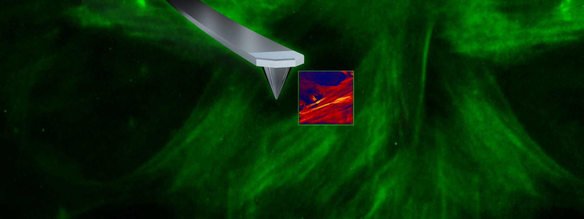
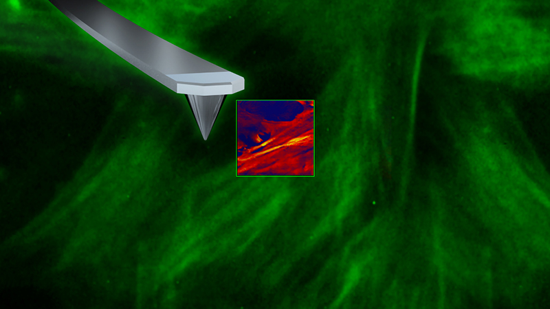
AFM-STED Correlative Microscopy
Claudio Canale, Ph.D., Department of Physics, University of Genoa, Italy
ABSTRACT: AFM-STED correlative microscopy was presented for the first time in 2012. In the last decade, this integrated technology has been employed to overcome some of the limits of single technique analysis. In this talk, the development of AFM-STED microscopy will be reviewed, starting from the motivation that drove the development of the first system to recent applications.
ABOUT THE PRESENTER: Dr. Claudio Canale, graduated in Physics, did his Ph.D. in Biophysics, and is currently a Senior Researcher at the University of Genoa, Italy. He began working on AFM in 2002 and has worked for many years on the study of the biological mechanisms associated with neurodegeneration in misfolding protein diseases. He contributed to the development of the first AFM-STED setup in 2012 while working at the Department of Biophysics of the Italian Institute of Technology.