Novel Electrical and Optical Neurotechnologies for Mapping Synaptic Integration Gradients in Vitro and in Vivo
Using electrical and optical neurotechnologies to explore dendritic spines.
Attendees will gain first-hand insights into how a combination of optical and electrical neurotechnologies provide novel insights into electrical transmission. Dr. Krishna Jayant discusses how he combines two-photon visualization with various techniques to perform:
- Targeted intracellular electrophysiology
- Simultaneous two-photon calcium imaging
- Stimulating spines in 3D
Webinar Summary
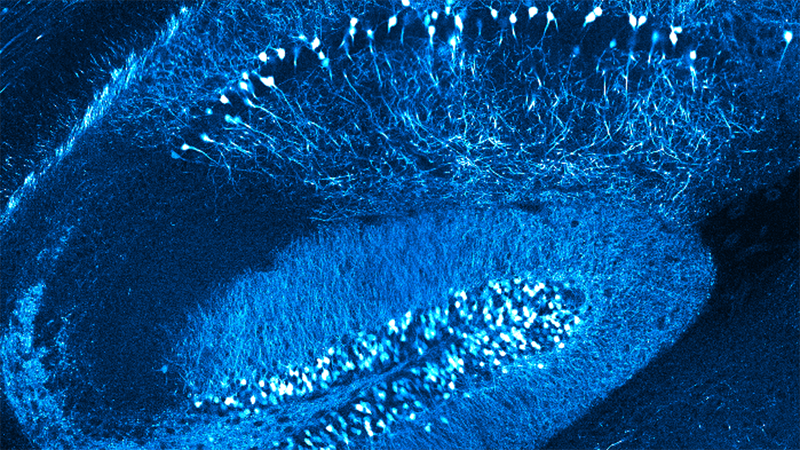
Dendritic spines, characterized by a small head (volume~0.01-0.1μm3) and narrow neck (diameter~0.1μm, length~1μm), are the primary site of excitatory synaptic input in the mammalian brain. Synaptic inputs made onto spines first integrate onto dendrites, and subsequently propagate towards the soma and axon initial segment, where they further integrate with other inputs to determine overall action potential output. Elucidating the electrical properties of spines is thus paramount for understanding the first steps along this signal-processing chain. Yet, their micron/sub-micron size has rendered conventional whole-cell intracellular electrophysiology infeasible.
In the first part of this talk, the speaker describes his work using quantum-dot labeled quartz nanopipettes (15-30 nm diameters) under two-photon visualization for targeted intracellular recordings from spines and small pre-synaptic terminals. He shows through detailed experiments that (i) spines receive large EPSPs (25-30mV), and (ii) estimated neck resistances are large enough to influence electrical isolation (mean ~420 MΩ) and filter synaptic input as it invades the dendrite. He briefly describes the theoretical implications of these properties.
In the second part of this talk, he describes a new method in which he combines the flexible property of these nanopipettes with microprisms to enable simultaneous two-photon calcium imaging and targeted intracellular electrophysiology across (a) cortical depth; (b) different cell types; (c) somatic and dendritic segments; and (d) anesthetized and awake head-fixed locomoting mice. As a prototypical application of the method, he describes targeted intracellular recordings from PV+ interneurons, while simultaneously imaging epileptic seizure spread across cortical layers. Dr. Jayant gives a succinct overview of recent work on biomimetic nanopipettes, custom CMOS intracellular amplifiers, and the fabrication of two-photon compatible electrode arrays. Finally, he concludes by describing recent endeavors from his lab at Purdue University on using two-photon holography to stimulate spines in 3D.
Find out more about the technology featured in this webinar or our other solutions for Super-Resolution Microscopy:
Speaker

Dr. Krishna Jayant
Assistant Professor of Biomedical Engineering, Purdue University, USA
Krishna Jayant received his B.Tech degree in electrical engineering from the National Institute of Technology (NIT) Tiruchirappalli, India in 2005, where as part of his bachelor’s thesis he worked on bio-inspired optimization techniques. After brief research stints at IISc Bangalore (2005-2006), and the University of Bologna (2006-2007), both in the area of microelectronics, he joined Cornell University, Ithaca NY, where he received his M.S/PhD in electrical engineering in 2014 working with Prof. Edwin C. Kan on Bioelectronics. He then joined Columbia University for postdoctoral research training with Profs. Rafael Yuste, Ken Shepard, and Ozgur Sahin in the field of circuit neuroscience, biophysics, and CMOS integrated neurotechnology. Here, he was awarded the prestigious Kavli Post-Doctoral Fellowship for carrying out work at the intersection of nanoscience and neuroscience. As an Assistant Professor at Purdue, his laboratory focuses on elucidating the biophysical features involved in neural computation and their behavioral relevance using a combination of nano-electrophysiology, optical microscopy, CMOS integrated systems, and theory. His research is supported by the NIH Trailblazer Award and the HFSP young investigator grant.