▶ Watch On-Demand | 60 Minutes

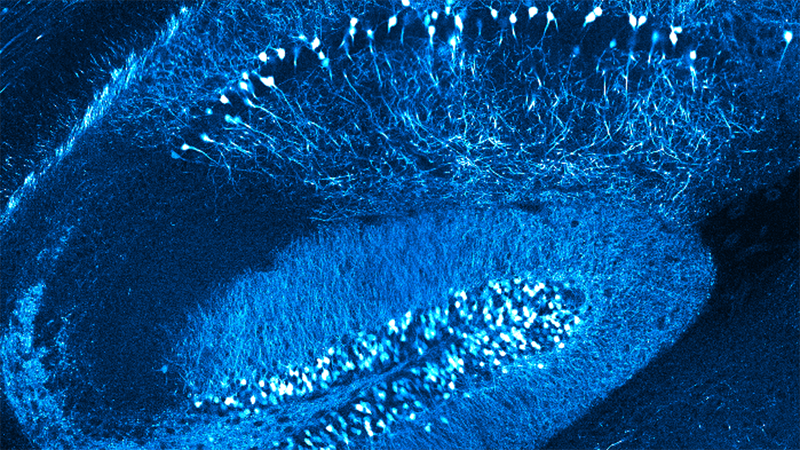
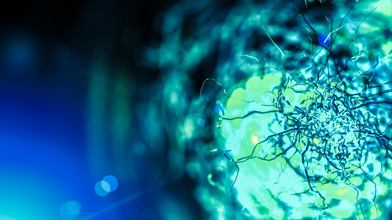
In Vivo Imaging of Microglia as the Brain’s Thermostat for Tuning Neuronal Activity

Dr. Mario Merlini presents new insights about using in-vivo two-photon imaging with calcium uncaging for the study of microglial regulation of neuronal activity
Presented by Dr. Mario Merlini, Junior Team Leader at the Blood & Brain Department, Caen-Normandie Institute (June 17, 2021)


PRESENTATION HIGHLIGHTS

  • [00:03:50] Introduction to the study of microglial regulation of neuronal activity
  • [00:06:30] Review of laboratory data
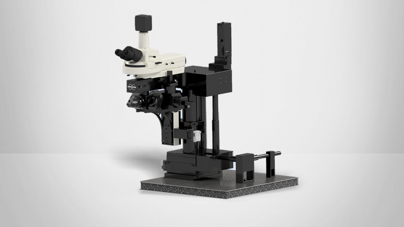
  • [00:11:30] In-depth presentation of two-photon imaging methods used in this study
  • [00:28:30] Main findings: Why do microglia dynamics respond and adapt to neuronal activity?
  • [00:34:00] Live question & answer session with the presenter

Expert Answers to Audience Questions

Q: How did you apply each glutamate? Specifically, how did you know that the concentration was correct, how did you ensure that the photoactivation was working, and how did you approach other elements of the experimental design?


Q: Did you perform imaging in both active (awake) and inactive (anesthetised) mice?


Q: In the microglia extensions and velocity analysis, how were you able to distinguish between processes as microglia tend to intertwine often?


Q: What was the promoter for the jRCaMP1b?


Q: Can you comment on your choice of using RuBi-Glutamate vs. MNI-glutamate?
Q: Can you provide more details about laser ablation and how you did it or this study?


Q: Can you explain why you chose to stimulate the whiskers while imaging moving mice instead of having immobilized mice and/or no stimulation?


Q: Microglia could get activated in the process of the cranial window and show more qualities similar to what you describe. Is this something you observed in your experiments and, if so, how did you deal with it?


Q: Do you have any preferences for microglia to target specific domains of the neuron (i.e. dendrites, cell body, etc.)?


Q: How does microglia send glutamate - is it MUR-dependent and what type of MUR is expressing microglia?
Q: Would you recommend any ranges for the laser power applied to the brain for glutamate uncaging?


Q: Do you have any thoughts on which of the GTPases was primarily responsible for skewing microglia dynamics in the PTX mice or do they all need to be activated?


Q: How did you correct the movement of the mouse during awake behavior when you were imaging the fine processes?


Q: How many lasers are on the instrument used in your experiments?