Tech Note: Supervised Feedback Microscopy with the Acquifer IM and the Plate-Viewer Software
Semi-automated approach for complex high-content screening experiments
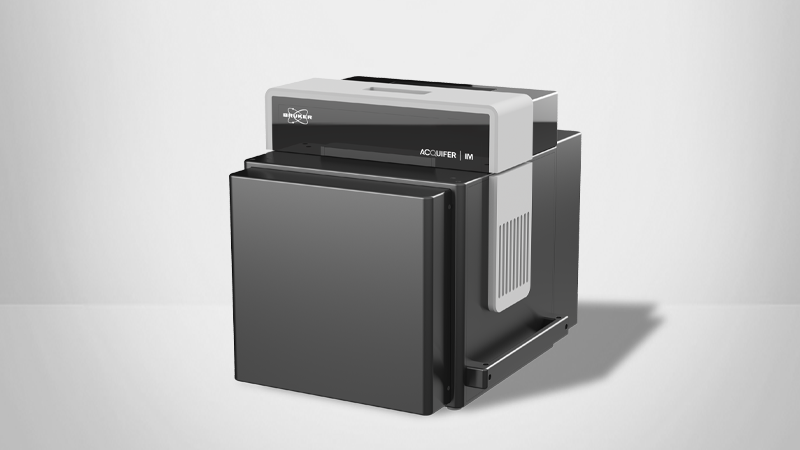
In this technical note, readers will learn about how the Acquifer IM is an ideal solution for advanced supervised feedback microscopy experiments including tissue-specific imaging of structures in large biological specimens. Unique features of the Plate-Viewer software on the Acquifer IM high-throughput screening microsope include the ability to select regions of interest automatically or to preview fields of view with an adjustable bounding box, which enables complex high-content screening experiments.
Readers can expect to learn more about:
- The Acquifer IM and the Plate-Viewer software address the limitations of traditional high-content screening microscopes
- A semi-automated approach is an ideal solution for supervised feedback microscopy
- User-friendly features of Acquifer IM enable efficient high-resolution data acquisition and visualization
- Tissue-specific imaging provides unique insights into the biological processes of large specimens
KEYWORDS: Acquifer Imaging Machine (IM), High-Content Screening, Supervised Feedback Microscopy, Semi-Automated Experiments, Plate-Viewer Software
Introduction
Modern high-content screening microscopes allow rapid automated imaging of entire microtiter plates by imaging fixed positions within each well. This is ideal for in-vitro cell culture-based readouts or other assays with evenly distributed phenotypes. However, it imposes limitations when large specimens or rare events are studied because the limited field of view (FOV) of high-magnification objectives may not encompass the full region of interest. Therefore, users are often limited to lower magnification acquisition, leading to low-resolution data, or omitting features of interest in many wells. This technical note discusses how Bruker's Acquifer Imaging Machine (IM) overcomes these challenges with the Plate-Viewer software, and offers a semi-automated approach for advanced supervised feedback microscopy experiments.
Automated Experimental Approaches
For tissue-specific imaging in large specimens (e.g., zebrafish) an approach is needed that can automatically identify and zoom in on the tissue or organ of interest. Fully automated tissue detection and imaging often demand the development of sophisticated image processing routines. Therefore, each project needs careful balancing between software development time and project size. Semi-automated approaches offer an ideal compromise, as they only require minimal user interaction and no custom algorithm development. Even complex or variable structures that would require extensive development of image detection routines can be readily handled via this simpler alternative. This approach is beneficial for screening projects as they start instantly, saving time and resources.
The Plate-Viewer Software on the Acquifer IM High-throughput Screening Microscope
Plate-Viewer software offers a semi-automated approach for supervised feedback microscopy. Low-magnification pre-screen data of a full microtiter plate is visualized following the plate layout for a quick and intuitive overview. The integrated “click-tool” functionality allows assay experts to select regions of interest (ROIs) for each well. Moreover, built-in and generic “template matching” algorithms enable robust automatic localization of various target structures. These can be complex reporter expression patterns, morphological features, or rare events in each well. Acquifer IM automatically acquires data at high resolution from selected regions according to predefined settings and acquired high-resolution data is then readily and intuitively visualized in the Plate-Viewer software.
Plate-Viewer has several unique features that enable researchers to:
- Select ROIs to be automatically imaged by a simple mouse click or template matching;
- Preview FOVs of higher magnification objectives using an adjustable bounding box;
- Intuitively visualize and browse even large-scale screening datasets;
- Readily navigate through complex multidimensional datasets;
- Apply LUTs and overlay channels for improved manual inspection;
- Adjust basic image parameters to enhance visualization of details; and
- Plug-in interface for integration of external image processing tools.
Intuitive Solution for High-Content Screening
Acquifer IM is a complete solution for supervised feedback microscopy experiments. The Plate-Viewer software on the Acquifer IM high-throughput screening microscope utilizes a semi-automated approach to greatly simplify complex high-content screening experiments. This innovative tool enables researchers to more easily and reliably perform tissue-specific imaging of complex or variable structures in large specimens.
©2024 Bruker Corporation. Acquifer IM and Plate-Viewer are trademarks of Bruker. All other trademarks are the property of their respective companies. All rights reserved. TN2604, Rev. A0.

