High-Speed AFM Imaging
High-Resolution Imaging of DNA

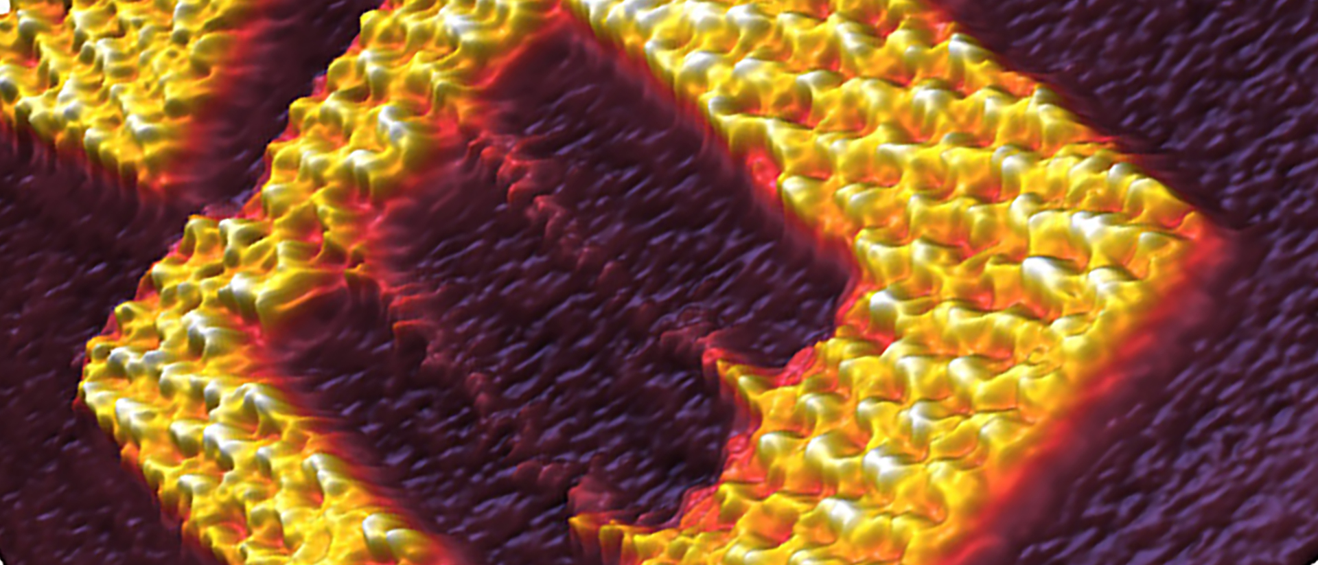
DNA Origami at 150 Lines/Sec
Tailored DNA origami frames imaged in TAE 10 mM MgCl2 buffer on mica with the JPK NanoWIzard BioScience 4 XP AFM. Scan field: 125 nm; height range: 4.4 nm; scan speed: 150 lines/sec.
Courtesy of:
R. Willaert, VUB
Brussels (BE)
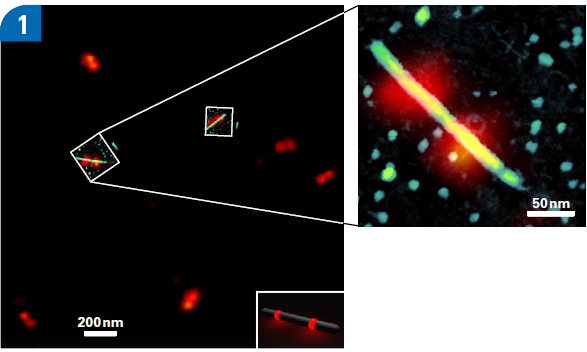
Correlative STED and AFM Images of Nanorulers
JPK NanoWizard ULTRA Speed images of a Correlative Experiment of AFM and STED on ATTO-647N-labeled DNA nanorulers in TAE-buffer. STED images show dimers of 12-15 ATTO-647N molecules 70 nm apart (see schematic inset). AFM QI image shows a 200 nm long DNA nanorod with 8nm diameter (height range: 12 nm).
Courtesy of:
GATTAQUANT GmbH (Germany)
VIDEO – Soft DNA Origami in Buffer
JPK NanoWizard ULTRA Speed video of the central pattern of a soft DNA Origami imaged in buffer. It shows 14 consecutive frames with a true line-rate of 105 Hz (256 pixels x 256 pixels, 0.41 frames per second). X,y - range 50 nm x 50 nm; z-range 600 pm. Cantilever Nanoworld USC-F0.3-k0.3.
DNA origami samples prepared as described by:
Sacca et al., Angew. Chem. Int. Ed. (2010) 9378
Samples provided by:
Dr. Rebecca Meyer, Prof. Christof M. Niemeyer
Karlsruhe Institute of Technology (KIT)
VIDEO – Streptavidin Binding to Biotinylated DNA Origami at 50 Frames and 5000 Lines/Sec
High-Speed AFM imaging in closed-loop with the JPK NanoRacer® of DNA origami nanostructures containing 5 biotin binding sites on mica imaged in buffer with streptavidin presence. Streptavidin binding/unbinding can be observed on top of the DNA origami nanostructures as bright dots appear/disappear. The video is taken at 50 frames/sec with true 5000 lines/sec and consists of over 1400 frames; X,Y-range 150 nm.
In collaboration with:
C.M. Domínguez, C.M. Niemeyer
Institute for Biological Interfaces (IBG-1)
KIT (Germany)
VIDEO – High-Speed Imaging of DNA at 10 Frames/Sec
JPK NanoWizard ULTRA Speed AFM images of the same individual DNA molecule acquired in liquid over 400 consecutive scans, which demonstrate the low invasiveness and stability (rate 10 frames/sec). With conventional AFM (4 lines/sec), this experiment would take more than 2 hours.
Live Cell Dynamics
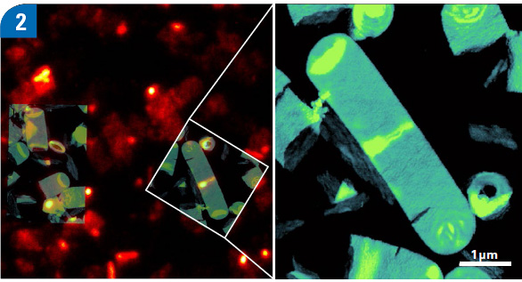
Correlative STED and AFM Images of Isolated Sacculi of Bacillus subtilis
Correlative STED and AFM images of isolated sacculi of Bacillus subtilis with cell division protein (FtsZ) labeled for highlighting z rings, captured on the JPK NanoWizard ULTRA Speed. AFM image in PBS with QITM (height range: 150nm).
Courtesy of:
R.K. Tank1,3 · R.D. Turner2,3 · S. Kumar1,3 N. Mullin1,3 · A. Cadby1,3 · S.J. Foster2,3 · J.K. Hobbs1,3
1 Department of Physics and Astronomy
2 Department of Molecular Biology and Biotechnology
3 The Krebs Institute; all University of Sheffield, UK
VIDEO – Live CHO Cell Dynamics on a Living Chinese Hamster Ovary (CHO) Cell
JPK NanoWizard 4 BioScience AFM video of a living Chinese hamster ovary (CHO) cell imaged in medium at 37°C. The unassisted video acquisition, consisting of 600 consecutive AFM phase images taken with a line-rate of 48 Hz (256 pixels x 256 pixels, tip velocity of 600 µm/s) depicts very diverse cell surface dynamics, involving cytoskeleton reorganization, as well as plausible membrane events. X,Y - range 5 µm x 5 µm.
Cantilever Nanoworld USC-F0.3-k0.3
Courtesy of:
The lab of Prof. Andreas Herrmann
Humboldt University
Berlin (Germany)
VIDEO – Living KPG7 Fibroblast Imaged in Cell Medium at 37 °C
JPK NanoWizard ULTRA Speed video of a living KPG7 fibroblast imaged in cell medium at 37 °C. The video consisting of 20 consecutive AFM phase images with a line-rate of 30 Hz (256 pixels x 256 pixels, ~ 8.5 s/frame) depicts the dynamic morphological changes taking place on the surface and the periphery of the cells. X,y - range 5 µm x 5 µm and z-range 6 degrees.
Cantilever Nanoworld USC-F0.3-k0.3
KPG7 fibroblasts, cultured in tissue culture dishes, provided by:
Dr. Roland Schwarzer, Prof. Andreas Herrmann
Humboldt University
Berlin (Germany)
Biodegradable Polymer Crystallization
VIDEO – Melting and Crystallization of a PCL Thin Film
JPK NanoWizard ULTRA Speed AFM images of a thin film of biodegradable polyester polycaprolactone (PCL) during melting and crystallization while ramping the sample temperature from 33°C to 62°C and down again. The NestedScanner technology allows high-speed scanning of 225 µm/sec during the entire temperature cycle while following the variation of the thickness of the PCL film (2.5 µm) during swelling and contraction. With this new technology, challenging samples with heights of up to 8 µm can, for the first time, be examined at the highest scan speeds.
- Left: Topography z-range 60 nm
- Right: Phase z-range 25 degrees
VIDEO – PHB/V Spherulite Crystallization
This JPK NanoWizard ULTRA Speed video shows the growth front of a polyhydroxybutyrate-co-valerate (PHB/V) spherulite crystallization (24x). It consists of 58 consecutive phase images with a true line rate of 60 Hz (512 pixels x 512 pixels) and 1.5 µm in x and y and a z-range of 19 degrees.
Courtesy of:
Prof. Jamie Hobbs
University of Sheffield
UK