Quantitative and Fast AFM Co-Localized with STED Microscopy in Living Cell Experiments
Open Up an Almost-Unlimited Array of Applications
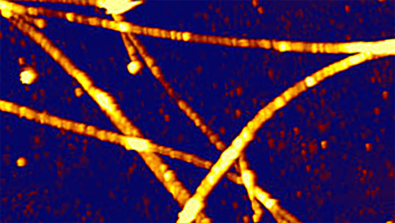
KEYWORDS: Corrleative Nanoscopy; Microtubules; STED; Super-Resolution
Integrating AFM with optical microscopy opens up new ways to study biological samples under physiological conditions, particularly when specificity is required. One popular correlative approach for nanoscale imaging of specifically labeled structures is the combination of AFM and super-resolution light microscopy. One of many super-resolution microscopy techniques used in these experiments stimulated emission depletion (STED).
A combined AFM and STED system is a powerful tool for biological applications. STED provides specificity, while AFM delivers true 3D surface visualization and nanomechanical information.
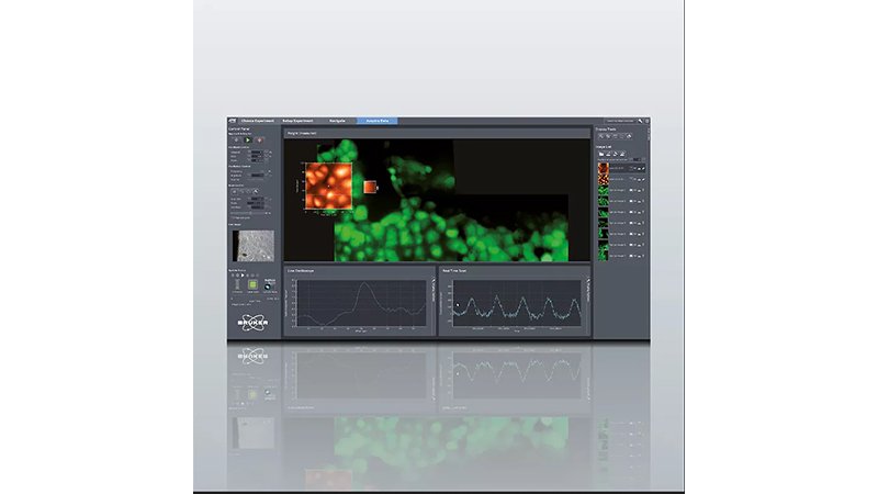
This application note showcases the performance of simultaneous STED and AFM measurements in nanomanipulation experiments. The measurements depicted are the first examples of an almost unlimited array of applications for the new and innovative correlative technique.
Readers can expect to learn about:
- The individual limitations and drawbacks of AFM and STED;
- How combining these techniques creates a more complete dataset and provides deeper insight into the specimen under investigation; and
- The use of STED and AFM to collect and correlate chemical and nanomechanical information, intracellular features, and topography.