EPR in Life Science
Membrane proteins and spin labelling
Mobility analysis of nitroxides in spin labeled protein
EPR in conjunction with site-directed spin labeling (SDSL) is a technique to study structure and dynamics of membrane proteins. EPR provides information about the local environment of the spin label that has unpaired electrons but also can measure inter-spin label distances when two spin labels are introduced inside the protein.
Metalloproteins
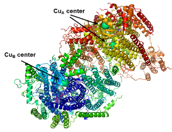
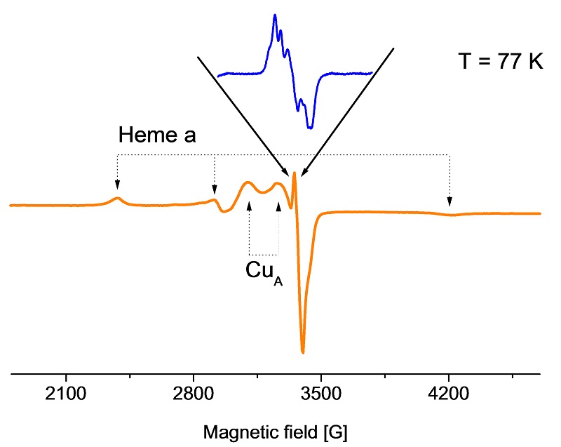
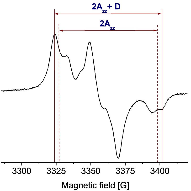
EPR characterization of paramagnetic centers in cytochrome c oxidase
Approximately 30% of all known proteins are metalloproteins. They are involved in a variety of biologically important processes such as electron transfer, drug metabolism, disease mechanisms, etc. EPR has an important role not only to study the electronic structure of metalloproteins but also to characterize their redox cofactors, binding sites, substrate reactions. For example, cytochrome c oxidase is a terminal protein in the respiratory chains from mitochondria and many bacteria. A low-spin heme, heme a, accepts electrons from a copper A (CuA) center bound to subunit II, and transfers them to a binuclear center.
Enzyme Reactions
Detection and study of the active site of Cu,Zn-SOD
Many enzyme reactions involve one-electron oxidation steps with formation of paramagnetic transient state of the enzyme detectable by EPR. The paramagnetic center where the unpaired electron is located, is usually centered at a transition metal (metalloproteins) or is an amino acid derived radical. Detection and identification of the paramagnetic centers is important to understand the function of the enzymes. For example, in the native SOD1 enzyme, the active site contains one Cu(II) ion that gives a very characteristic EPR spectrum.
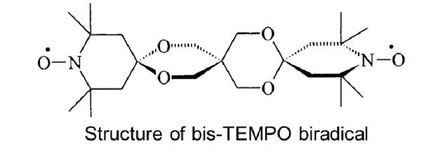
Screening DNP agents
EPR spectrum and dipolar coupling determination of bis-TEMPO
Correct concentration of DNP polarizing agents is crucial to the success of a DNP experiment. Samples can be pre-screened before DNP experiments using the patented SpinCount module, even in the MAS rotor. Relaxation times are critical for DNP efficiency therefore P1/2 measurements at low temperature to estimate the DNP efficiencies of new polarization agents are invaluable. Another characteristic of importance in DNP measurements is the electron-electron dipolar coupling that is easily measured from solution and frozen solution EPR spectra.
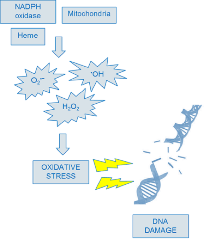
RNA and DNA oxidation
DNA-derived radicals detected upon CuCl2/H2O2 treatment
EPR spectroscopy in conjunction with spin trapping has been employed successfully to detect and identify high-molecular-weight species generated as a result of reactive oxygen species (ROS)-induced damage to biological macromolecules, such as DNAs and RNAs. The destruction or alteration of these materials is known to play a key role in a large number of cellular injuries and diseases.
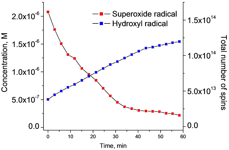
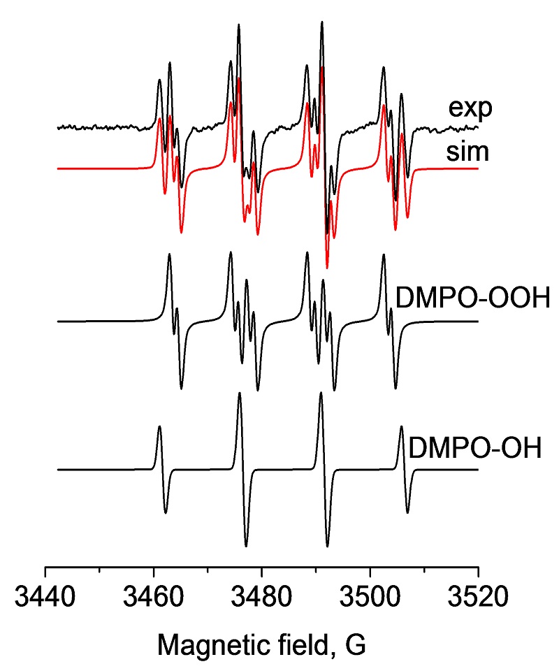
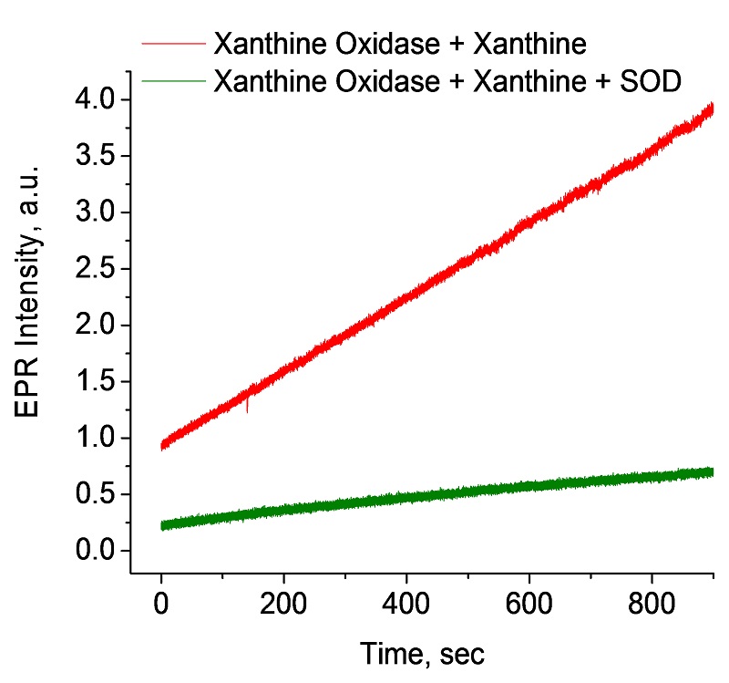
Detection of Reactive Oxygen Species (ROS) using spin traps
Quantitative EPR analysis of superoxide and hydroxyl radicals
Oxidative stress and damage in cells is associated with the development of cancer, Alzheimer‘s disease, atherosclerosis, autism, infections and Parkinson‘s disease. Reactive Oxygen Species (ROSs) are the main cause of oxidative stress and damage in cells, causing damage to proteins, lipids and DNA. Two leading ROS are radicals such as the superoxide radical (O2•-) and the hydroxyl radical (HO•) as shown here in the Xanthine/Xanthine oxidase system where their generation and decomposition can be accurately followed with the EMXnano.
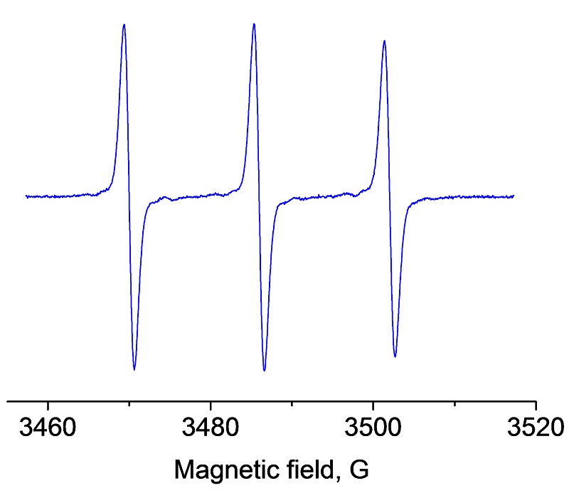
Detection of Reactive Oxygen Species (ROS) using spin probes
Time course of superoxide formation using the spin probe CMH
In vascular cells, increased generation of superoxide (O2•-) has been suggested to occur in hypertension, diabetes, and heart failure. Thus the accurate detection and ability to quantify O2•- are critically important in understanding the pathogenesis of these various cardiovascular disorders and other noncardiovascular diseases. As shown here the generation of superoxide over time can be easily monitored with the EMXnano.
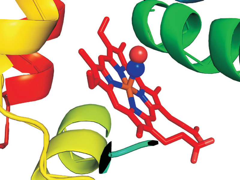
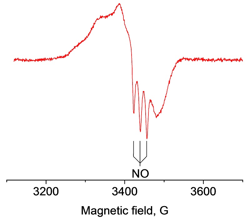
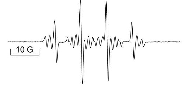
Nitric oxide
Binding of nitric oxide to oxyhemoglobin detected at 100 K
Nitric Oxide (NO) is a highly reactive regulatory molecule which has many important physiological roles, such as a neurotransmitter in the central nervous system, a regulator of vasomotor tone in the cardiovascular system, and a cytotoxic mediator of the immune system. NO is a free radical and its short half-life (< 30 sec), has rendered direct measurement difficult. The instability of NO can be overcome by using a NO-trapping technique, in which a more stable complex is formed and subsequently detected by EPR. For example, the oxidation of nitric oxide (NO) to nitrate by oxyhemoglobin (oxyHb) is a fundamental reaction in NO biology and binding of NO to the heme can be characterized by EPR.