CUBES
Modular Benchtop Imaging Solutions for preclinical PET, SPECT and CT imaging research.
Intuitive workflows, high performance and seamless multimodality made accessible to every lab.
Scalable Nuclear Imaging Platforms
Compact in size
CUBES: Nuclear Molecular Benchtop Imaging
At Bruker, we are committed to making nuclear molecular imaging accessible to every research lab. The CUBES enable researchers to visualize and quantify biological processes in vivo using high-performance PET, SPECT and CT imaging. These compact and modular devices lower the barrier to preclinical PET, SPECT and CT imaging while delivering the highest performance, supported by fast and simple turnkey workflows.
CUBES are designed for use as stand-alone units and in multimodal combinations, offering seamless and fully integrated multimodal nuclear imaging workflows from a single User Interface and precise co-registration between all modalities.
The benchtop design and a plug-and-play architecture allow you to combine any number or combination of PET, SPECT and CT CUBES at any time, so you can scale your research capacity at you own pace.
With over 250 CUBES installed worldwide, Bruker is your trusted partner in advancing preclinical imaging.
Features
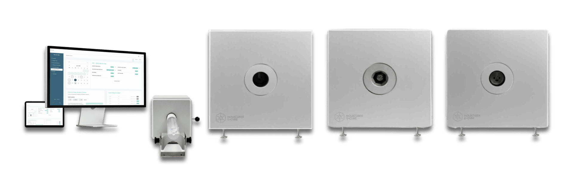
CUBES imaging platform
The CUBES imaging platform includes everything researchers require to run any combination of PET, SPECT or CT CUBES. It includes our intuitive CUBEFLOW software platform on workstation & tablet, one set of animal cradles for mice and rats, the animal preparation station and an anaesthesia scavenging system.
The CUBEFLOW software environment controls and monitors all CUBES from a single intuitive user interface on your workstation or tablet.
- Intuitive planning and reproducible setup of longitudinal studies
- Easy Scan mode
- Image acquisition and reconstruction on individual CUBES
- Animal monitoring on all CUBES
- Automated and periodic advanced quality control platform
Animal Cradles and Accessories
CUBES in vivo animal cradles for mice and rats are designed for stable physiological conditions under anesthesia during imaging.
- Range of animal cradles for single- or multi-animal imaging
- Fully integrated animal monitoring
- Automated supply and extraction of anesthesia gas
- Feedback regulated body temperature control and warming
- Auto-detection of animal cradles by each CUBE
The animal preparation station facilitates preparation of the animal of before imaging. Integrated physiological monitoring, anaesthesia and animal warming assure full control and animal safety.
The BioShield Mouse cradle provides a controlled pressure conditions, supporting studies that require enhanced biosafety conditions.
ZeroShift cradles allow transfer of anesthetized animals between all Bruker preclinical imaging modalities without changing the animal position, enabling a seamless co-registration of imaging results from MRI, PET, SPECT, CT, BLI and FLI.
PET Imaging: the β-CUBE
The β-CUBE enables sub-milimeter, quantitative in vivo PET imaging for non-invasive, longitudinal studies of tracer biodistribution, binding affinity, pharmaco-kinetics, time-activity characteristics or more.
- Static and dynamic imaging of up to 4 mice simultaneouly
- Cardiac gated imaging
- Image analysis and kinetic modelling with Bruker pmod
Characteristics:
- Transaxial FOV: 72 mm
- Axial FOV: > 250 mm
- Best Spatial Resolution: < 700 um
- Best Sensitivity >12%
- SiPM / LYSO digital PET detector
- Full detector-based DOI resolution
- weight / size: ~90kg / 54x56x54 cm

SPECT Imaging: the γ-CUBE
The γ-CUBE facilitates accurate, quantitative in vivo SPECT imaging. Reliable imaging of high-energy theranostic radionuclides, dynamic imaging protocols and detection of small or low-uptake biological signals are enabled, supporting the needs of any preclinical research lab.
- Multi-isotope SPECT imaging up to 511 keV
- Static and dynamic imaging of up to 3 mice simultaneouly
- Cardiac imaging
- Image analysis and kinetic modelling with Bruker pmod
Characteristics:
- Transaxial FOV: up to 60 mm
- Axial FOV: > 250 mm
- Best Spatial Resolution: 500 um
- Best Sensitivity: >1.2%
- Energy range: up to 511 keV
- Energy Resolution: <10%
- Patented ultra-lofthole collimators
- SiPM / NaI(TI) detector technology
- weight / size: ~130kg / 54x56x54 cm
CT Imaging: the X-CUBE
The X-CUBE combines fast whole-body CT imaging at extremely low dose with excellent soft tissue contrast for X-ray imaging research, morphological reference imaging and quantitative PET and SPECT image reconstruction.
- Low dose imaging of up to 4 mice simultaneouly
- Cardiac and respiration gated imaging
- X-ray dose estimation
Characteristics:
- Gantry: 86 mm
- Transaxial FOV: 65 mm
- Axial FOV: > 250 mm
- Spatial Resolution: 50 um
- 20-50 keV
- Fully X-ray shielded
- weight / size: ~120kg / 54x56x71 cm