Fluorescence Microscopy Upgrade
Fluorescence guided nanomechanical and nanotribological characterization
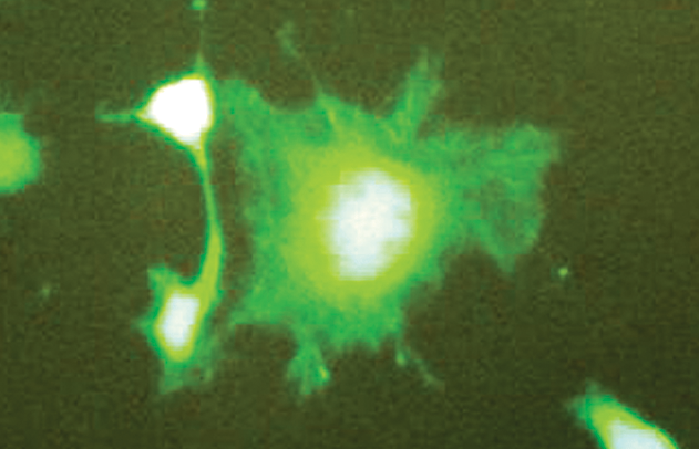
Fluorescence Microscopy combined with nanomechanical testing provides the ability to accurately position nanomechanical tests on specific regions of material marked with fluorochromes. Common bright field microscopy, standard on most nanomechanical test instruments, utilizes broad-spectrum reflected light to image the sample surface and cannot readily distinguish testing regions of interest on complex inhomogeneous materials. By allowing researchers to selectively tag individual constituents within the material with fluorochromes, accurate and reliable test positioning can be achieved on the region of interest.
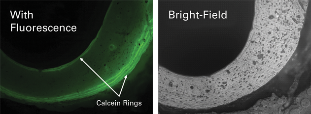
Target specific components of inhomogeneous materials
Bruker has developed a fully integrated fluorescence microscope for TriboIndenter Series Instruments. Nanoindentation and fluorescence imaging is combined utilizing a high-precision automated staging system and provides exceptional spatial synchronicity between the techniques. The fluorescence microscope utilizes user-definable filter sets for use with a wide range of fluorescent dyes and is also capable of bright field imaging, allowing the nanoindenter system to be effectively utilized in a variety of research areas.