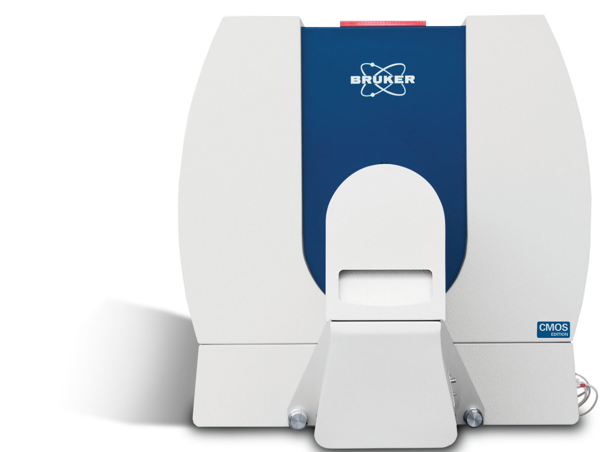
SKYSCAN 1276 CMOS EDITION
and soft tissues in small laboratory animals and biological samples
Highest resolution desktop
in vivo microCT
Highlights
The SKYSCAN 1276 CMOS Edition is a high performance, stand-alone, fast, desktop in vivo microCT with continuously variable magnification for scanning small laboratory animals (mice, rats, ...) and biological samples. An optional Large Animal Transport System (LATS) is available which enables the system to mount also large animals including rabbits.
The SKYSCAN 1276 CMOS Edition has an unrivalled combination of high resolution, big image size, round and spiral (helical) scanning and reconstruction, and low dose imaging. The image field of view up to 75 mm wide and 310 mm long allows full body mouse and rat scanning. The variable magnification allows scanning bone and tissue samples with high spatial resolution down to 2.8 µm pixel size. Variable X-Ray energy combined with a range of filters ensures optimal image quality for diverse research applications from lung tissue to bone with metal implants. The system can perform scanning with continuous gantry rotation and in step-and-shoot mode with scanning cycles down to 3.9 sec. Furthermore, the SKYSCAN 1276 CMOS in vivo microCT administers a low radiation dose to the animals allowing multiple scans in longitudinal preclinical studies without the risk of unwanted radiation-induced side effects. The fully integrated physiological monitoring package allows monitoring and controlling the animal's wellbeing at all times through a video stream, ECG, temperature and breathing detection.
The SKYSCAN 1276 CMOS is complemented by 3D.SUITE. This extensive software suite covers GPU-accelerated reconstruction, 2D/ 3D morphological analysis, as well as surface and volume rendering visualization.
Features
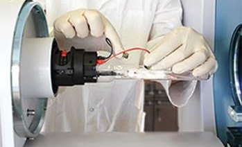
Mouse and Rat Cassettes
The SKYSCAN 1276 CMOS system is supplied with exchangeable animal cassettes that can be used in all Bruker in-vivo imaging instruments such as MRI, micro-PET, micro-SPECT, bio-luminescence, biofluorescence, etc. to collect multimodal information. It allows co-registration of functional and morphological information from the same animal.
Touchscreen Control
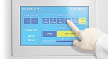
The user interface of the SKYSCAN 1276 CMOS system is simple and intuitive. The instrument can be controlled from the computer screen and also from the embedded force-sensitive touchscreen, which can be operated by gloved hands. The touchscreen allows selection of scanning protocol, adjusting the animal bed position and control of imaging and scanning. Where multiple scans are started from the touchscreen, the software will automatically save acquired data to separate subfolders with incrementally assigned folder names and dataset file prefixes.
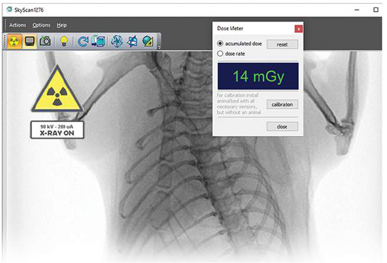
ON-SCREEN REAL-TIME DOSE METER
The SkyScan1276 control software includes a real-time on-screen dose meter. It indicates an estimation of the dose absorbed by the animal body during scanning. The measurement is based on the absorption calculated from X-ray projection images of the animal cross-calibrated with electronic dosimeter measurements. The dose meter shows accumulated dose or dose rate. It is calibrated for X-ray absorption in the standard mouse and rat cassettes. In this way it measures the X-ray dose absorbed in animal body itself during scanning. The dose absorbed by the animal during a scan is documented in the scan log-file together with all scan and reconstruction settings.
Integrated physiological monitoring
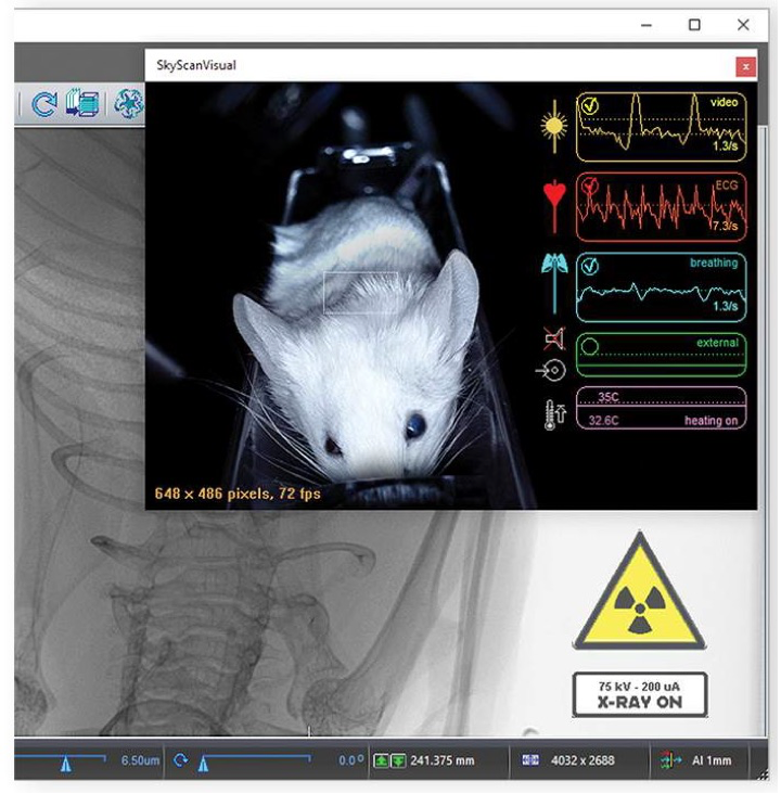
The physiological monitoring system includes video monitoring of an animal with real-time movement detection, ECG and breathing detection, and temperature stabilization. A 5 megapixel color camera is mounted above the animal bed along with white LED illumination to introduce a real-time image of the animal during the scan. The software analyses the video stream from a user-selected area of the image where breathing movement is visible. These movements are converted into a movement waveform to provide breathing time marks for time-resolved reconstructions. The face mask on the animal bed is connected to an air/gas flow sensor for direct breathing detection. The ECG electrodes in the animal cassette are connected to a sensitive ECG amplifier. Both breathing and ECG signals are digitized and displayed as real-time profiles on-screen. The monitoring also includes temperature stabilization by heated airflow, which maintains the scanned animal at a selected temperature, to prevent cooling of the animal under anaesthesia.
Highlighted Applications
Mineralized Tissues

Bruker’s third generation SKYSCAN 1276 CMOS is – it’s fair to say – the leading in vivo microCT bone solution, proving its worth in hundreds of research animal facilities worldwide. No other system has an x-ray camera optically tailored precisely for that impossible combination – high resolution with low dose, for the rodent bone. The full range of rodent and small animal bone disease models can be assessed and quantified reliably for study after study.
- Physiological monitoring is design-central in SKYSCAN in vivo scanners, not an add-on. High quality video, breathing monitoring by video-movement and pressure, ECG and thermostat-controlled warming, put the animal’s welfare at center stage.
- A large carbon fiber bed for rats and a steel tray for rabbits, for orthopedic implantation studies. Optimal hindlimb positioning for bone scans in mouse, rat and rabbit.
- Bone morphometry (ASBMR nomenclature) with comprehensive 3D and 2D parameters, with densitometry including BMD calibration references over a 2-32mm diameter size range.
- Advanced functions include 3D registration for longitudinal in vivo studies, adaptive thresholding, Euler connectivity, fractal, anisotropy and stereology, filtering, Boolean logical operators and much more.
Pneumology
The SKYSCAN 1276 CMOS is a flexible high-performance lung imaging solution. Sharply time-gated lung images are obtained in short scan times with safe levels of ionizing radiation. In the same scanner there is ex vivo sample scanning capability at a voxel size down to 2.8 micron true resolution allowing imaging lung tissue at the alveolar level of architecture for advanced lung disease characterization.
- Physiological monitoring is design-central in SKYSCAN in vivo scanners, not an add-on. High quality video, breathing monitoring by video-movement and pressure, ECG and thermostat-controlled warming, support in vivo lung studies with minimized welfare impact on the animal.
- Advanced animal cassettes connected for full physiological monitoring and anesthesia yet detachable with a single locking button – the universal Bruker in vivo bed. Support for third party devices such as ventilators (e.g. Scireq FlexiventTM).
- Comprehensive time-gating solutions: prospective and retrospective, time and image based intrinsic gating. Continuous rotation available for fast low-dose gated scanning.
- Full in-house software solution for lung function analysis in 4D: tidal volume, lung volume and HU density for pathology assessment, lung tumour morphometric segmentation, 3D registration of sequential scans to map disease progression, auto-separation of lung from other animal tissues in CT-Analyser software.
Cardiovascular Applications
Cardiac time-gated 4D imaging of the rodent is the technical benchmark of microCT imaging in vivo and Bruker’s SKYSCAN 1276 CMOS meets this challenge. Sharply time-resolved cardiac-lung single or dual-gated images are provided in short scan times with safe levels of ionizing radiation – reliably for study after study.
- Physiological monitoring is design-central in SKYSCAN in vivo scanners, not an add-on. High quality video, breathing monitoring by video-movement and pressure, ECG and thermostat-controlled warming, support in vivo cardiac-lung studies with minimized welfare impact on the animal
- Advanced animal cassettes connected for full physiological monitoring and anesthesia provision yet detachable with a single locking button – the universal Bruker in vivo bed. Support for third party devices such as ventilators (e.g. Scireq FlexiventTM).
- Comprehensive time-gating solutions: prospective and retrospective, time and image based intrinsic gating. Continuous rotation available for fast low-dose gated scanning.
- Full in-house software solution for cardiac function analysis in 4D: ejection volume, other cardiac chambers and aorta, time-analysis of cardiac wave-train including regularity and fractality, with flexible segmentation and analysis of the cardiac cycle in CT-Analyser software.
Application Gallery
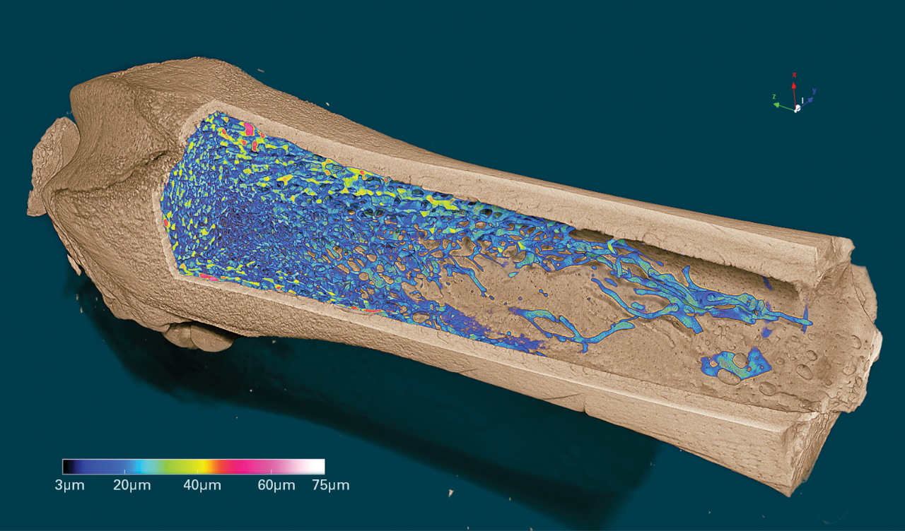
Volume rendered 3D model of a femur with color-coded representation of the trabecular thickness, scanned at 2.8 μm pixel size.
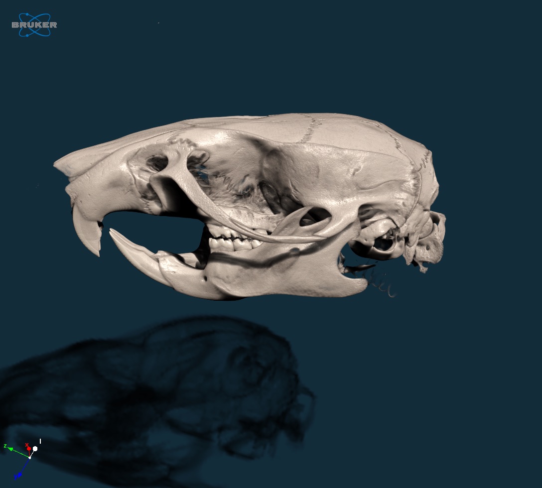
3D model of a rat skull, scanned at 20µm voxel size in vivo.
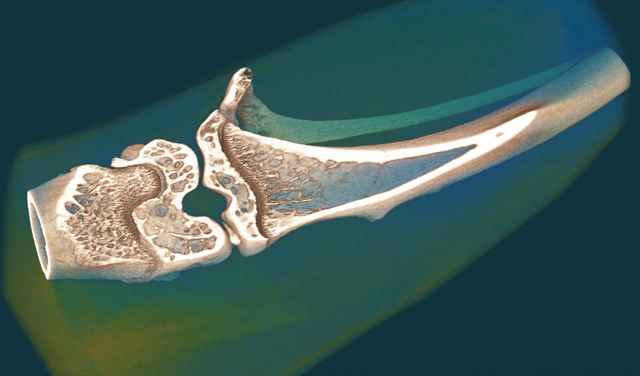
Volume rendered 3D model of a mouse knee, scanned at 6 µm voxel size in vivo.
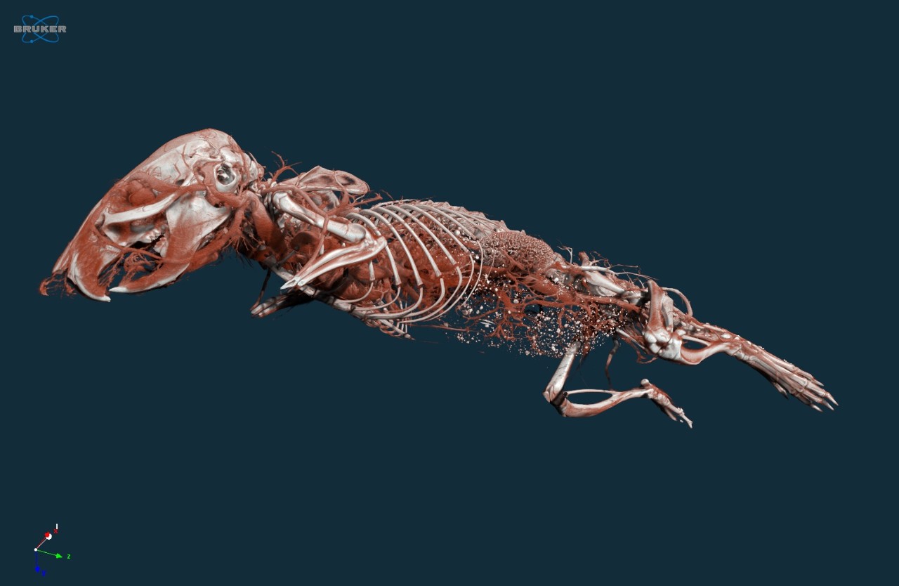
3D representation of the mouse vasculature, scanned in vivo at 7 µm voxel size after a bolus injection of vascular contrast agent.
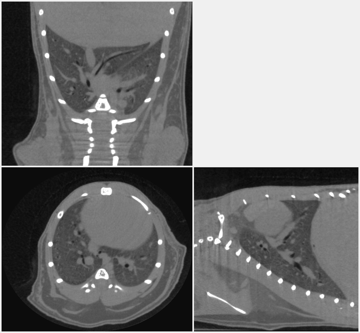
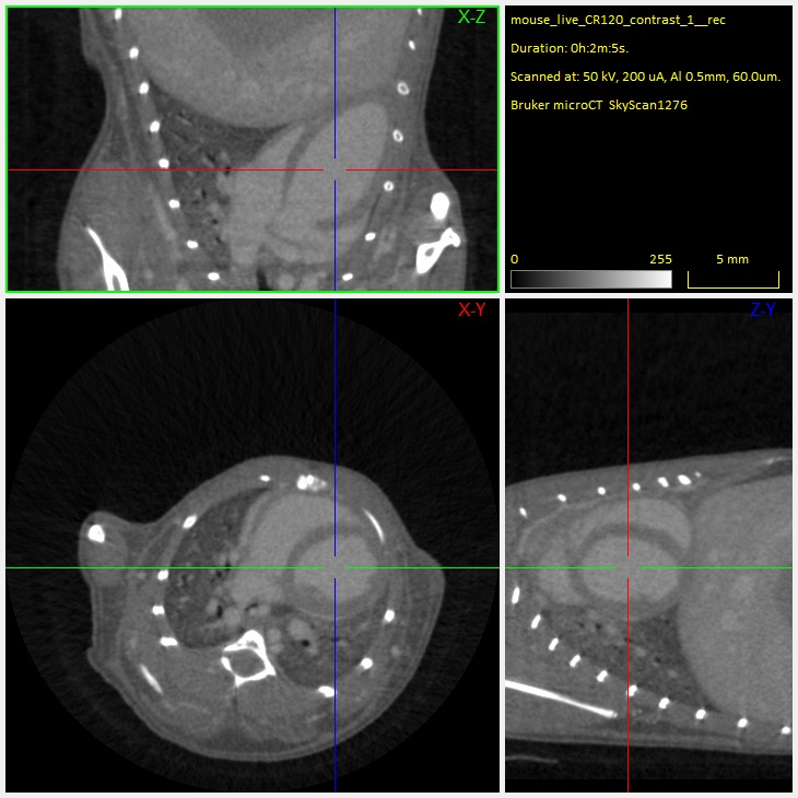
Orthogonal cross-sections through the mouse lung scanned in vivo, showing the blood vessels and large airways inside the lung.
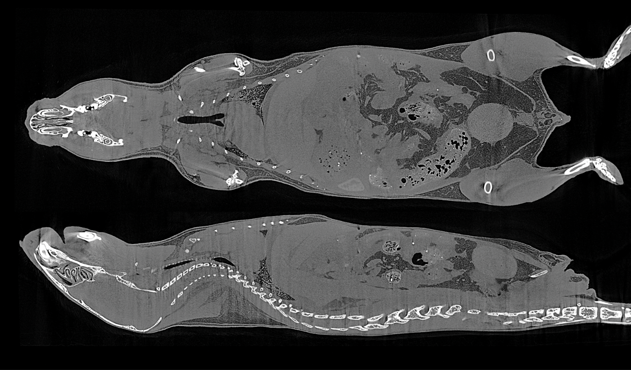
Cross-sectional slices through a mouse body, scanned at 17 µm voxel size in vivo without contrast agent injection.
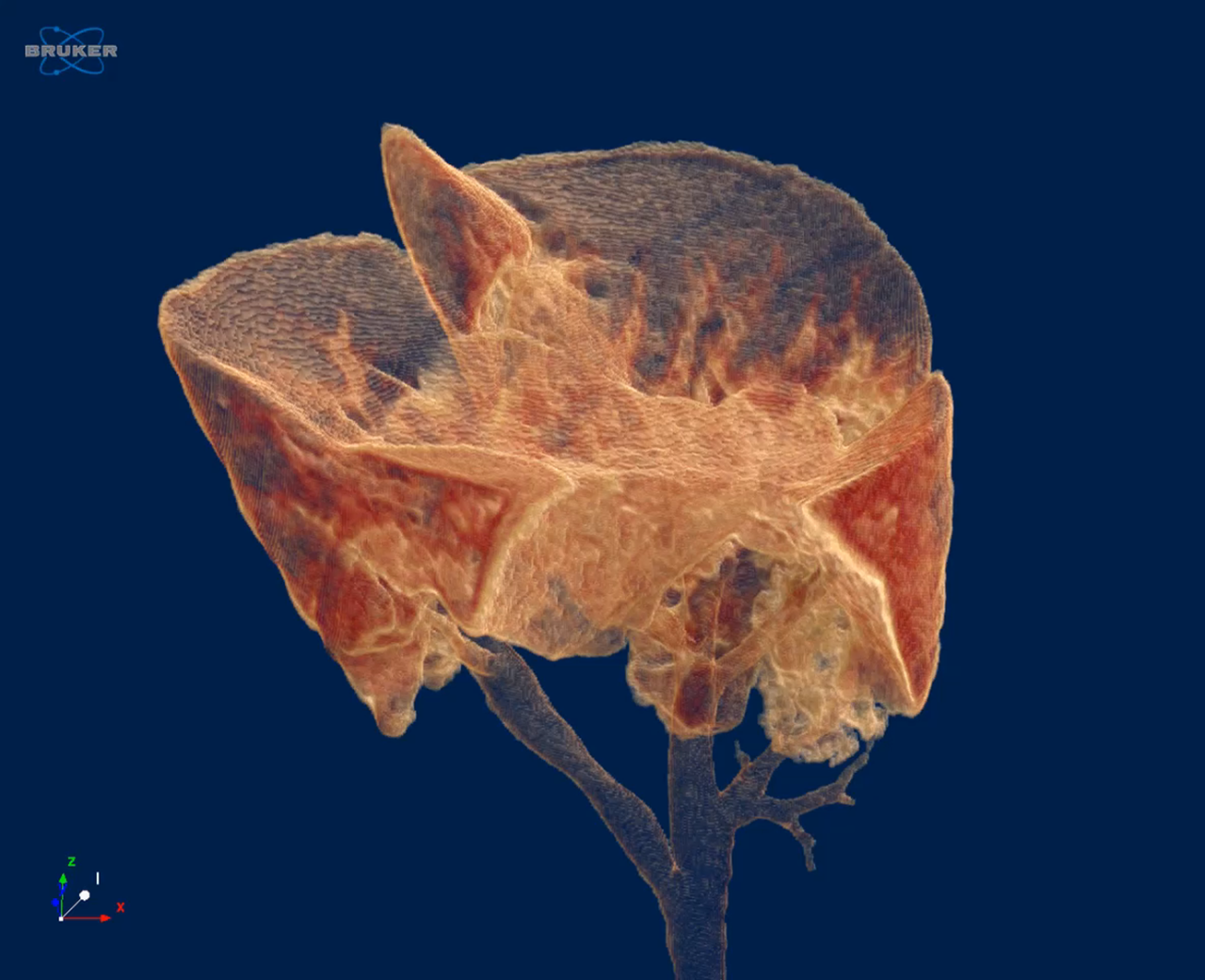
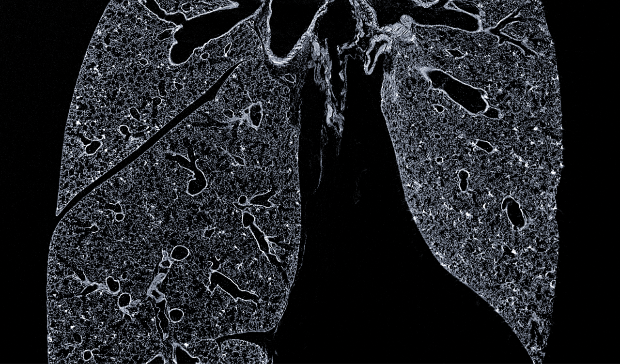
Cross-section through a mouse lung, scanned ex vivo after chemical drying at 3 µm voxel size.
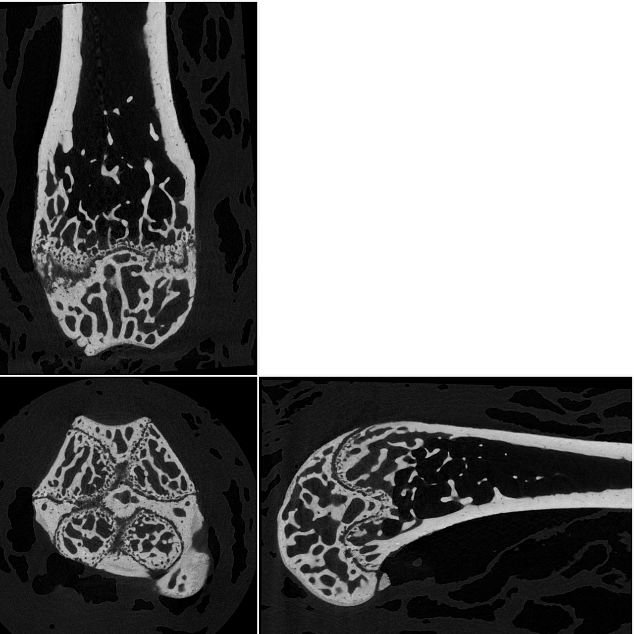
Orthogonal slices through a mouse femur, scanned at 2.8 µm voxel size.
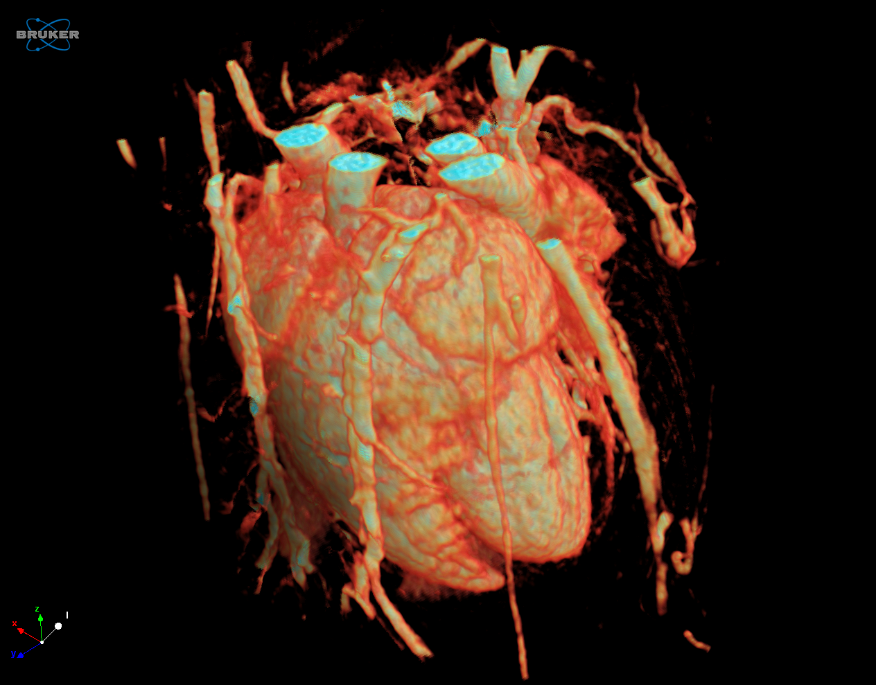
Orthogonal slices through a mouse heart, scanned in vivo after contrast agent injection.
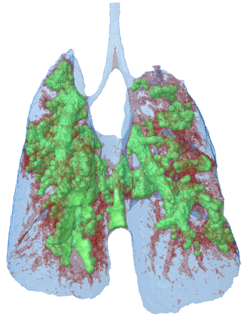
3D model of a mouse lung, made transparent to visualize the lung tumour tissue in green.
Specifications
SKYSCAN 1276 CMOS Specifications
Feature | Specification | Benefit |
X-ray source | 40 – 100 kV, 20W < 5 µm spot size at 4 W Automated 6-position filter changer | Maintenance-free sealed high resolution X-ray source |
X-ray detector | 16 Mp sCMOS detector (4096 x 4096 pixels) | Fine-pitched detectors for achieving highest resolution |
Spatial resolution | 2.8µm smallest pixel size 6µm spatial resolution (10% MTF) | Highest true resolution for in vivo imaging |
| Object size | 75 mm diameter 310 mm height | Capable to scan a large range of sample sizes |
Dimensions | with ATS: 954mm W x 940mm H x 1190mm L (1560mm L with open specimen chamber), 330kg with LATS: 954mm W x 940mm H x 1340mm L (1740mm L with open specimen chamber), 450kg | Space-saving desktop system that fits in every lab |
Power supply | 100-240V AC, 50-60Hz, 3A max. | Minimum installation requirements, a standard power supply suffices |
POSITION, SCAN, RECONSTRUCT and ANALYZE
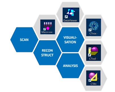
Bruker XRM solutions include all software needed to collect and analyze data. An intuitive graphical user interface with user guided parameter optimization support both expert and novice users. By using the latest GPU powered algorithms, reconstruction time is substantially reduced. CTVOX, CTAN and CTVOL combine to form a powerful suite of software for both qualitative and quantitative analysis of models.
3D.Suite Software:
Bruker microCT solutions include our comprehensive, in-house developed
3D.SUITE software for reconstruction, inspection, visualization, and
analysis of the internal object structure
Measurement Software:
SKYSCAN 2214 – Instrument control, measurement planning and collection
Reconstruction Software:
NRECON – Transforms the 2D projection images into 3D volumes
Analysis Software:
DATAVIEWER – Slice-by-slice inspection of 3D volumes and 2D/3D image registration
CTVOX – Realistic visualization by volume rendering
CTAN – 2D/3D image analysis & processing
CTVOL – Visualization of surface models to export for CAD or 3D printing
SKYSCAN 1276 - Service & Support
Available services include
- Help desk support from highly-skilled troubleshooting professionals, to isolate and resolve hard- and software problems
- Web-based remote instrument service for service diagnosis and applications support
- Merged reality support with Help Lightning – a virtual engineer at your side (video)
- Planned maintenance, according to your requirement
- Customer on-site repair and maintenance service
- Spare parts availability typically over night or within a few working days worldwide
- Compliance services for installation qualification, operational qualification / performance verification
- Site planning and relocation
Bruker Support
On our support website you will find:
Software Updates
- System software updates are available online to registered users.
Documentation
- Product manuals and installation guides are available online to registered users.
LabScape
Service & Life Cycle Support for Magnetic Resonance and Preclinical Imaging
Bruker’s commitment to provide customers with unparalleled help throughout the buying cycle, from initial inquiry to evaluation, installation, and the lifetime of the instrument is now characterized by the LabScape service concept.
LabScape Maintenance Agreements, On-Site On-Demand and Enhance Your Lab are designed to offer a new approach to maintenance and service for the modern laboratory