Intro
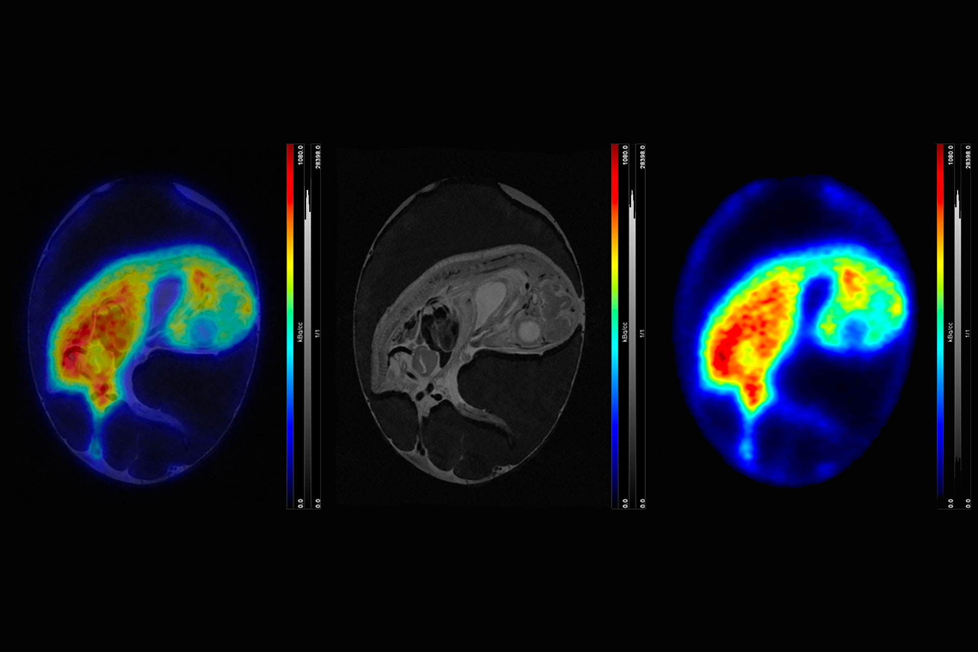
Despite great progress in the development of advanced in vitro models such as co-cultures, organ-on-a-chip, and microfluidic systems, in vitro models still lack the physiological complexity of a living organism regarding blood flow, metabolism, and endogenous membrane barriers. The use of fertilized chicken eggs (in ovo model) bridges the gap between cell-based assays and rodent models, rendering this model a useful tool for the initial evaluation of new radiotracers regarding their in vivo performance. Simultaneous PET/MRI offers great potential in terms of soft tissue contrast, as it enables clear differentiation between embryonic organs and tumor tissue, thus allowing very accurate assignment of the PET signal to embryonic structures.
Webinar Overview
The chick embryo and its chorioallantoic membrane (CAM) have been used in biomedical research for centuries, leading to fundamental knowledge about the developing chick embryo and groundbreaking discoveries in cardiovascular and tumor biology. This webinar will showcase different applications of this model in preclinical imaging. The contribution of the in ovo model to the 3R principle in tracer development will be discussed. The experimental workflow, starting with the incubation and handling of fertilized chicken eggs, intravenous injections into CAM-vessels, immobilization of chick embryos, image acquisition, and quantification, will be illustrated. Challenges and potential disadvantages will be discussed, and practical advice will be given.
Thursday, 20 November 2025
4 PM CET
What to Expect
The webinar will discuss the contribution of the in ovo model to the 3Rs principle in tracer development and will provide guidance on how to perform in ovo PET/MR imaging.
Key Learning Points
- Simultaneous PET/MR of chick embryo and its chorioallantoic membrane (CAM)
- Alternative imaging platform for faster & cost-effective tracer development
- Experimental procedures, advantages & challenges
Who Should Attend
This webinar is of interest to researchers who are developing new radiotracers or contrast agents and are looking for faster and more cost-effective methods besides wanting to explore alternatives to current animal models in the context of the 3Rs.