Three-Photon Imaging
In this video: Jimmy Fong (Director, Products and Technology, Bruker Fluorescence Microscopy) describes how Bruker’s three‑photon–compatible optics enable long‑wavelength imaging for experiments that require increased penetration depth. He highlights a thin‑skull imaging approach that leverages three‑photon excitation to visualize microglia–vascular interactions without performing a full craniotomy.
Example data demonstrate how large collection optics support clear cellular imaging several hundred microns through the skull, expanding the types of biological preparations that can be studied with three‑photon microscopy.
Contact us or continue reading to learn more.
Diving Deeper: Three-Photon Microscopy and Imaging
What is three-photon microscopy?
Three-photon microscopy is a fluorescence microscopy technique where, in contrast to two-photon microscopy, the fluorophore absorbs three photons almost simultaneously. These excitation photons have longer wavelengths and lower energy than those used to excite the same fluorophore in one- or two-photon excitation microscopy.
How do researchers benefit from using three-photon microscopy?
Two-photon excitation microscopy has facilitated fundamental discoveries in life science research. Unfortunately, two-photon technology suffers from some limitations. Specifically, when imaging at and below 500 µm, the background signal increases and the contrast decreases — making it impossible to resolve either structures or physiological activity. Conversely, the physical characteristics of the excitation light in three-photon excitation microscopy offer deeper imaging with superior confinement of the excitation, thereby providing better contrast and signal-to-background ratio (SBR) than two-photon microscopy.

Equipped for Advanced Applications
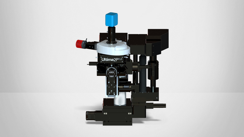
Bruker's Ultima 2Pplus multiphoton microscope is equipped standard to enable three-photon imaging experiments. Lens coatings throughout the optical path are designed for using extended wavelength laser sources, with special care taken in the design of the system to allow for short laser pulse-widths at the sample, which is critical for efficient three-photon excitation. Customers continue to successfully publish exciting three-photon experiments on Bruker multiphoton microscopes, and we hope to grow the community using this technique.
Practical Considerations for Three-Photon Microscopy
What are the limitations of three-photon microscopy?
The primary obstacle to the popularization of three-photon imaging has been the predicted increase in water absorption at the long wavelength window, as this effect might lead to overheating of the sample. However, researchers have found that the optimum wavelength for three-photon imaging is a trade-off between absorption and scattering in the 1300 nm and 1700 nm windows. Specifically, there is 2x more absorption but also almost 2x less scattering at 1300 nm, and as a result, imaging requires less laser power to be used. This demonstrates that there is enough room to accommodate research goals while complying with the physical constraints of three-photon microscopy. The convenience of three-photon imaging further balances out these constraints, as many common indicators used in two-photon microscopy can be easily re-purposed for use in three-photon imaging.
It is also important to consider the cost of the special laser sources and imaging lenses suitable for three-photon imaging. Typically, two lasers are needed for this application. To increase the probability of three-photon absorption, researchers must use a light source with a high photon density. The most common approach is based on a high-power laser pumping an optical parametric amplifier (OPA), which takes light of one wavelength from a femtosecond laser and turns it into light of two different wavelengths. The resulting beam, called the idler beam, that demonstrates a longer wavelength than the initial pumped laser is ideal for three-photon imaging. OPA is a low repetition laser (~1-4 MHz) and due to this characteristic, imaging with a resonant scanner at 30 Hz speed is not allowed. Typical imaging rates are less than 10 Hz with smaller fields-of-view, which could improve with the advancement of laser technology.