MALDI Imaging
MALDI Imaging - Reveal Greater Molecular Insight
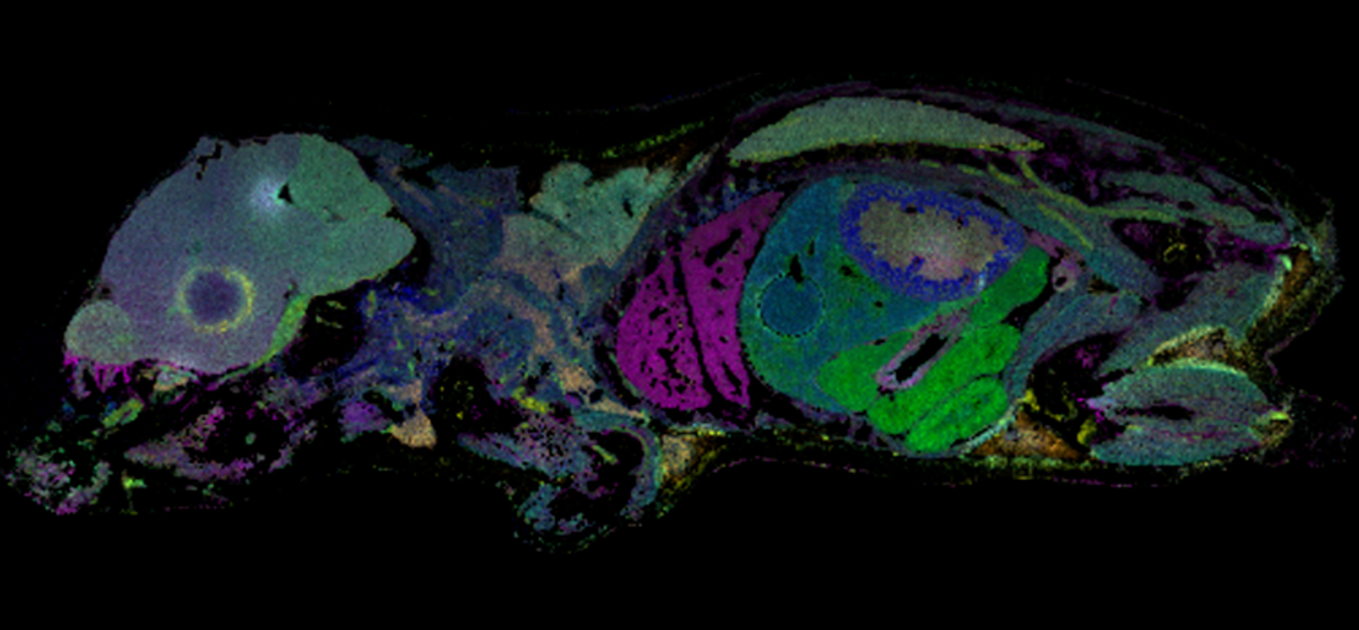
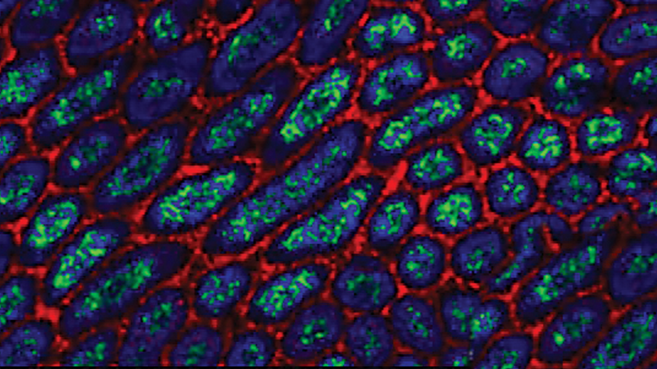
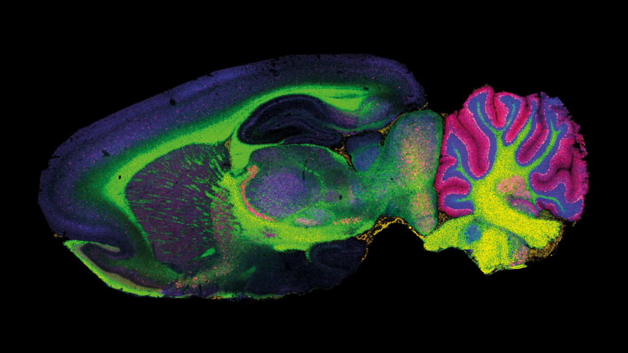
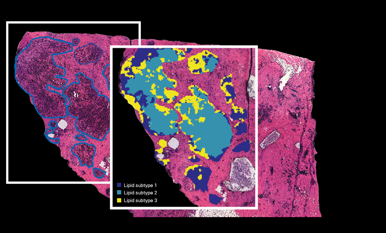
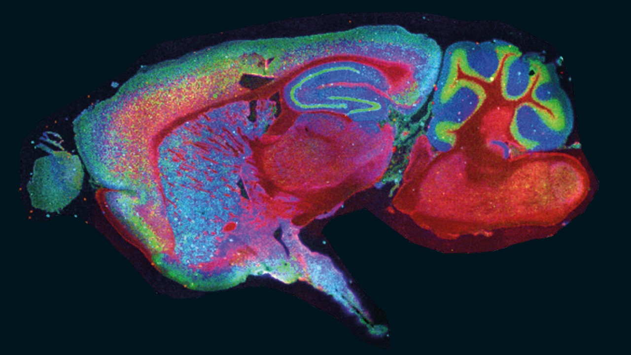
A single MALDI Imaging measurement produces up to several thousands of distribution maps, or ion images, to reveal greater insight and understanding of molecular makeup and regional heterogeneity.
- Label-free mapping of many analytes, metabolites and proteins
- Image hundreds to thousands of compounds
- Probe the molecular heterogeneity of samples
- Identify regionally specific molecular differences
- Seamless integration with microscopy or histology
Molecular Imaging Redefined
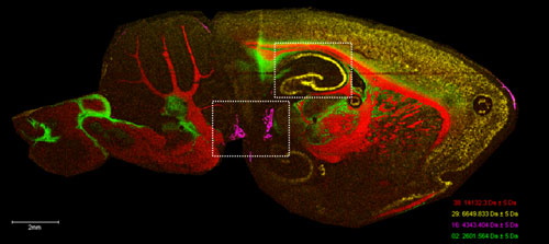
Whether your research is focused on plant metabolomics, biochemistry of cellular processes, biomarkers of disease development or treatment, or microbial interactions, MALDI Imaging can deliver new insight to your work. Key to this is the ability to integrate MALDI Imaging with other imaging modalities you may already be using, such as fluorescence microscopy or histology.
Bruker pioneered this capability in 2005 by allowing the importation and registration of other images into the MALDI Imaging dataset with full visual overlay.
Application areas for MALDI Imaging are diverse and growing, driven by the label-free nature of the technique and the ability to differentiate compounds by molecular weight. Untargeted discovery studies such as those in clinical research seek to uncover novel biomarkers of disease and treatment. For maximum information with intelligence use MALDI guided SpatialOMx®. MALDI Imaging is also revolutionizing pre-clinical drug discovery pipelines by providing direct distribution monitoring of therapeutic compounds and their metabolites.
These are two examples of untargeted or targeted methodologies where MALDI Imaging has proven valuable. Newer applications surrounding plants, polymers and microbes can be seen emerging in the scientific literature where Bruker's MALDI Imaging Technologies have proven valuable.
MALDI Imaging Technologies
Using MALDI to spatially measure molecular signatures
For more than 25 years, Bruker has been the leader in MALDI mass spectrometry innovation. MALDI is amenable for any analytical task and this is exemplified in the scientific literature where MALDI Imaging publications dominate 5:1 over the next ionization technology. Bruker delivers MALDI Imaging systems that incorporate novel MALDI technologies to accomplish any targeted or untargeted imaging study.
Spatially oriented molecular signatures
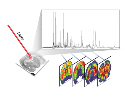
Images are generated by collecting MALDI data in a virtual grid pattern applied to a sample surface. Thin sections of samples are mounted onto conductive glass slides and a layer of MALDI matrix is applied by aerosol. Bruker’s MALDI Imaging technologies allow the user to define the measurement area and resolution directly using a digital image of the sample. The size of the virtual grid establishes the image resolution and is tailored for the experimental question to be answered.
Given each MALDI spectrum is acquired from a discrete virtual grid without oversampling, the resulting dataset is a molecular signature of the unique area sampled. Unlike ionization systems which utilize oversampling, Bruker’s MALDI Imaging technologies produce ion images which do not exhibit image ‘blurring’.
Imaging Technologies - Analytical Versatility and Imaging Speed
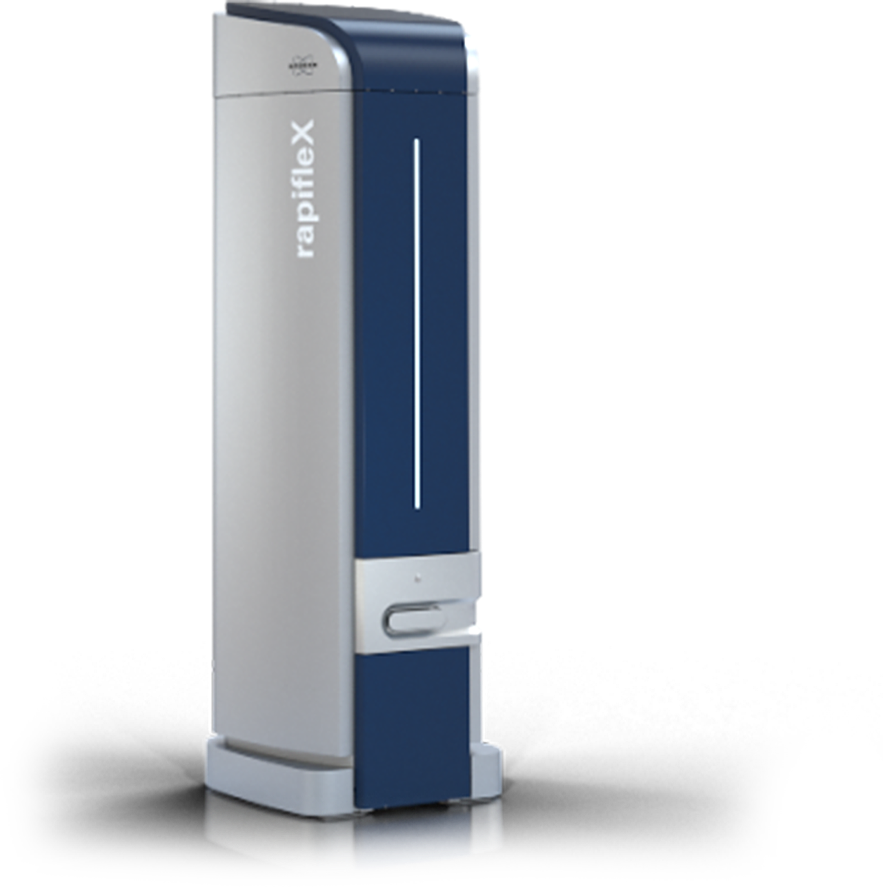
For biomarker discovery imaging studies, Bruker’s FLEX series of MALDI-TOF and TOF/TOF systems offer the greatest analytical versatility and imaging speed. For this reason, MALDI-TOF platforms are the most widely used imaging systems found in scientific literature.

Highest Molecular Specificity
Untargeted and targeted studies of smaller molecules demand high analytical performance for separating isobars and/or near-isobars. Bruker’s MRMS and timsTOF fleX systems meet the challenge by offering the highest molecular specificity available.
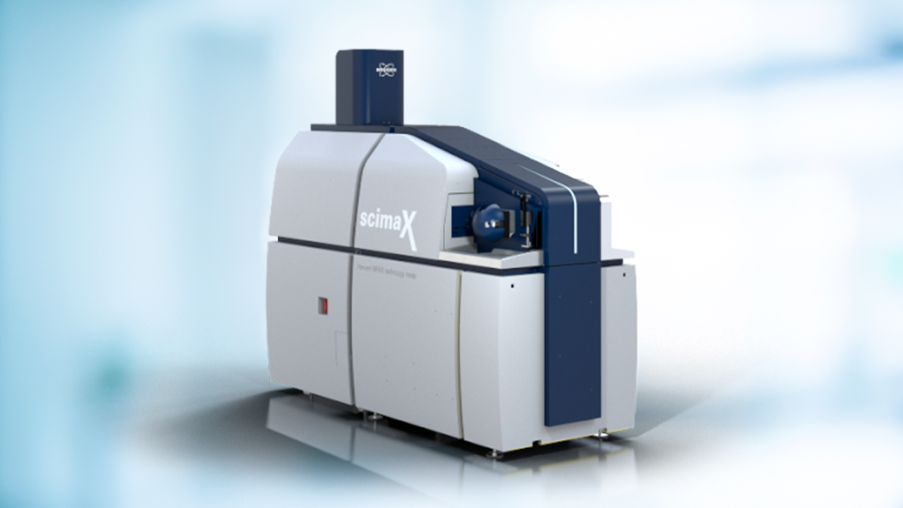
For targeted and untargeted imaging studies, Bruker’s solariX and scimaX® Magnetic Resonance Mass Spectrometers (MRMS) offer eXtreme mass resolution. At imaging speeds, achieve > 290,000 mass resolution at m/z 400 capable of differentiating and mapping compounds that differ by only a few mDa.
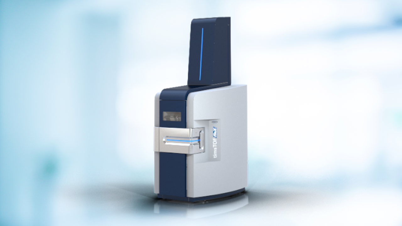
The timsTOF fleX offers versatility needed for SpatialOMx®. Built on Bruker’s pioneering timsTOF Pro platform with PASEF®, the timsTOF fleX powers all your multiomics analyses, while adding a high spatial resolution MALDI Imaging source to deliver a spatial dimension to OMICS analyses. Transform proteomics analysis into spatial proteomics, lipidomics into spatial lipidomics, and metabolomics into spatial metabolomics - SpatialOMx®.
SCiLS™ autopilot: Automated workflows to simplify measurement setups
Our software solutions make it easy to start with MALDI Imaging. The SCiLS™ autopilot e.g., is a new software workflow for simple and reproducible setup of MALDI Imaging measurements relying on Bruker’s IntelliSlides®. The automated steps save time and reduce manual user input increasing reliability and reproducibility of the measurement regardless of users’ expertise.
The automated workflows are available for the timsTOF fleX, timsTOF fleX MALDI-2 and rapifleX®.

For Research Use Only. Not for use in clinical diagnostic procedures.