MALDI HiPLEX-IHC
True Multiomics driven by MALDI HiPLEX-IHC
Discover the complex network of biological interactions that define biological processes from regulated homeostasis to disease pathology and progression within organs, tissues, and cellular systems.
Connect Cellular Signaling Pathways with Targeted Proteins through True Multiomic Imaging
Spatial localization of key protein expression targets is fundamental to the understanding and definition of disease, especially cancer biology, and single cell and bulk analysis for the characterization of cellular phenotypes.
Available tools for protein spatial profiling mostly come with limitations that can hinder building a complete picture of spatially defined biology such as intermediate plex levels that cannot track a sufficient number of proteins, limitide fields-of-view and inability to probe Omics layers outside of protein and RNA targets.
MALDI HiPLEX-IHC brings intact protein imaging to the next level by combining transcriptome relevant information from labelled protein expression mapping with advanced small molecule imaging from the same tissue section using MALDI Imaging. It is now possible to not only map proteins, but also examine metabolic activity and dig deeper into the biology governing cellular interactions, tissue homeostasis, and disease progression.
A new technology for spatial biology Imaging
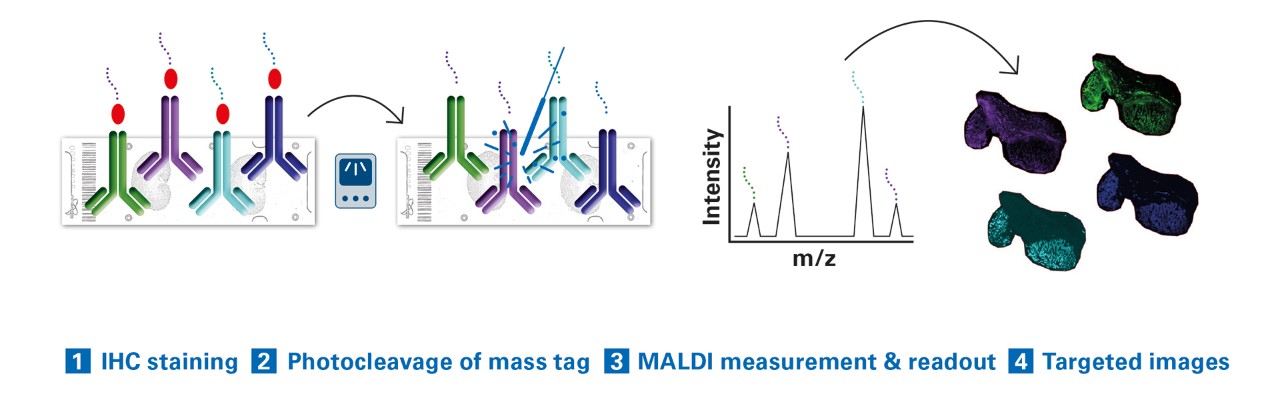
Multiplex. 100+ biomarkers, single scan, no cycling. Every probe comes with a unique mass tag.
Multimodal. Fluorescence microscopy, histopathological staining, and MALDI Imaging on one tissue section.
Multiomic. One workflow to visualize small molecules, lipids, glycans, intact proteins, and other macromolecules.
Fast and complete information. Survey an entire slide in 40 min. Drill down to ROIs at 5 µm high resolution with microGRID.
Easy Workflow. No conjugation. Routine IHC staining. No metal tags.
Whole slide imaging. No compromises on what part of the tissue section to image.
How it works
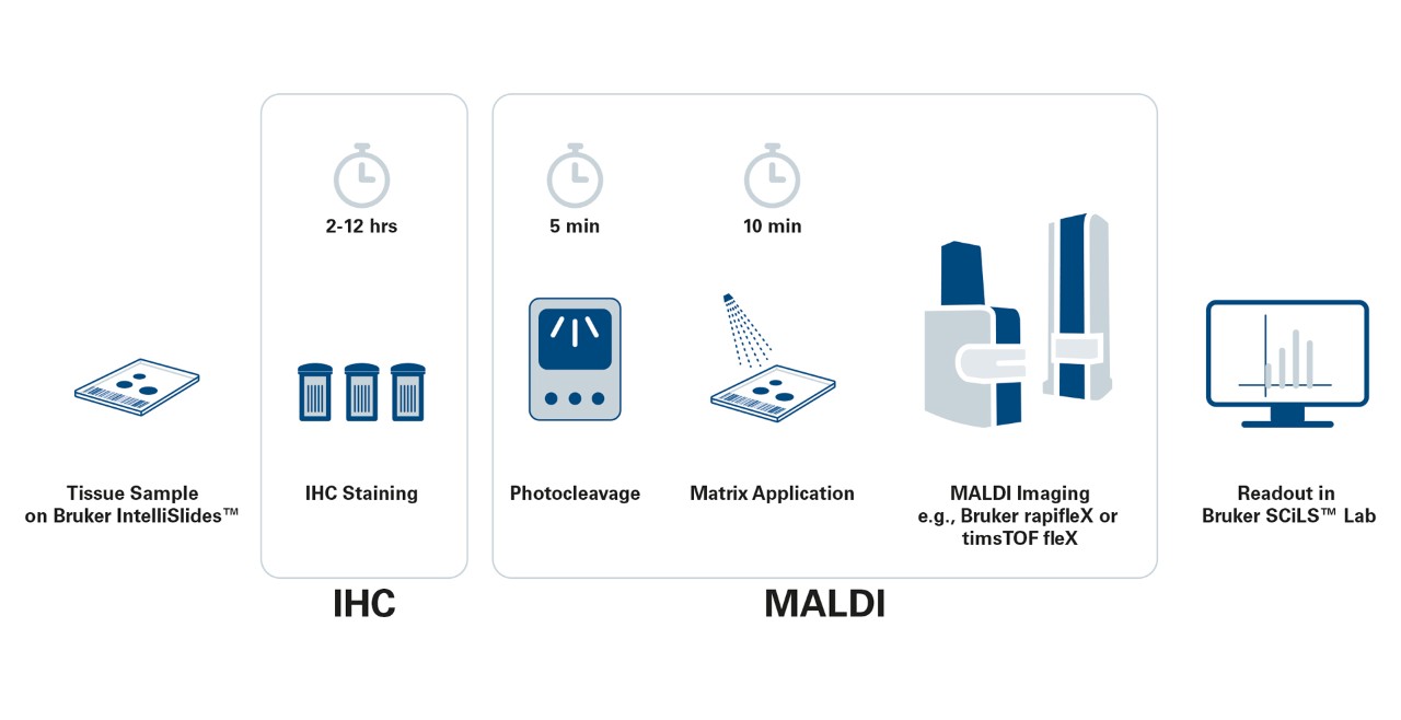
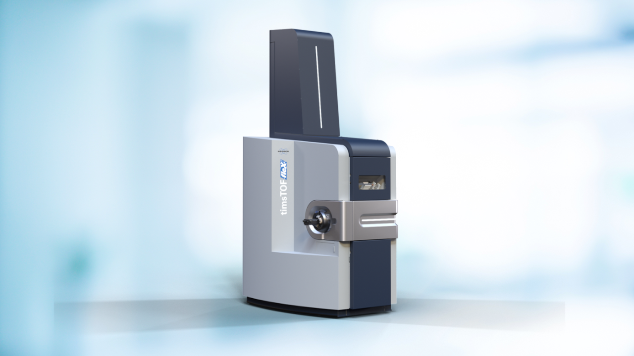
MALDI HiPLEX-IHC fits seamlessly into existing MALDI workflows for Bruker’s proven timsTOF fleX and rapifleX® platforms.
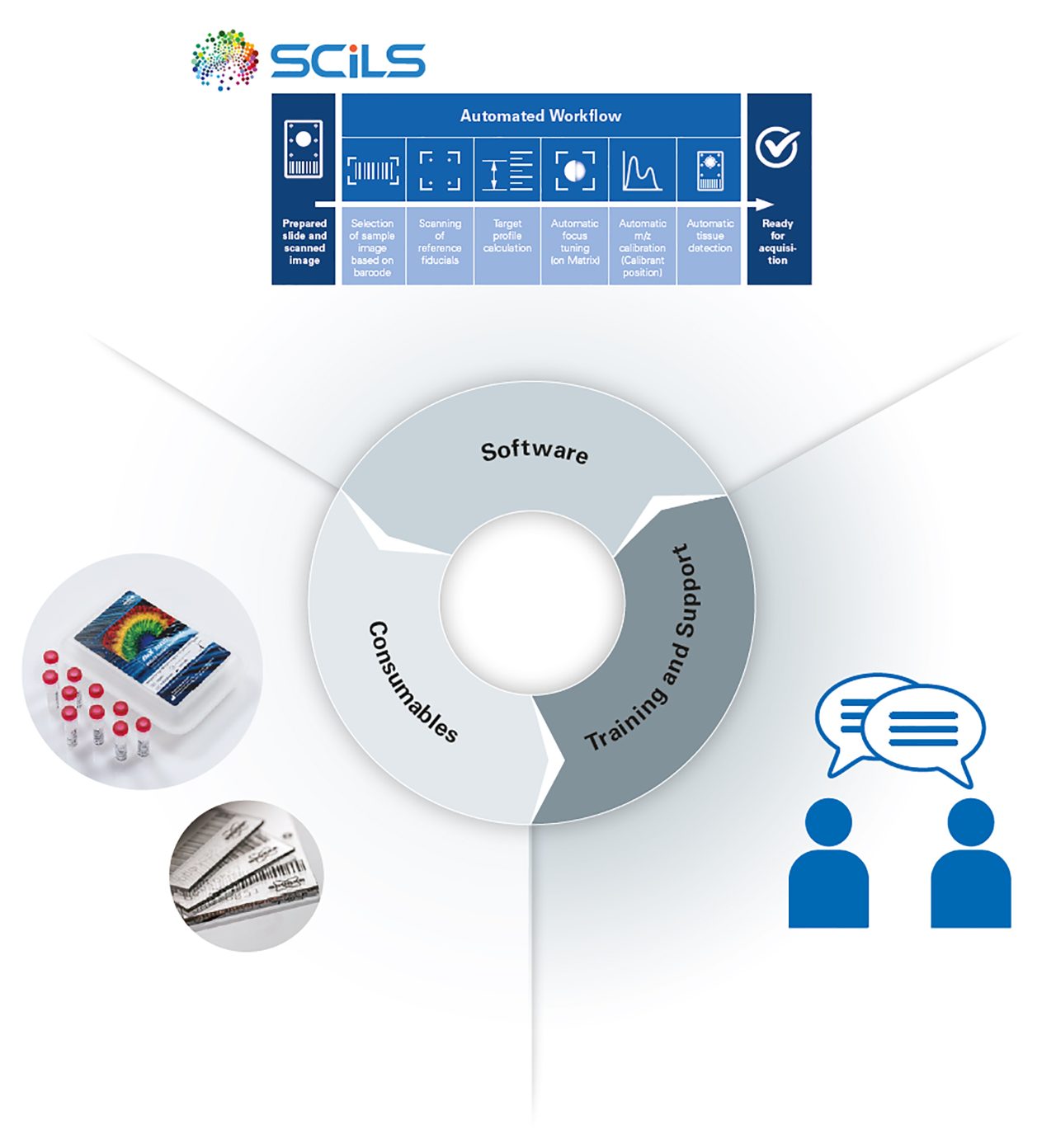
Full software automation through SCiLS™ autopilot allows for “pushbutton” workflow setup, ensuring reproducible and consistent preparation without the need for highly trained experts.
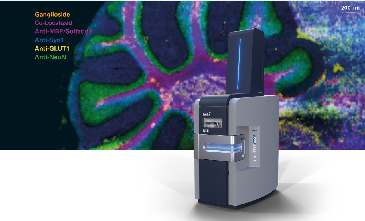
timsTOF fleX – Complete multiomics platform with spatial contextualization
- Visualize maximum biological information from proteins, metabolites, lipids, and xenobiotics
- Uniquely ID non-targeted small molecules with CCS databases or link to LC-MS/MS
- MALDI-2 adds wide chemical dimension to the small molecule overlay
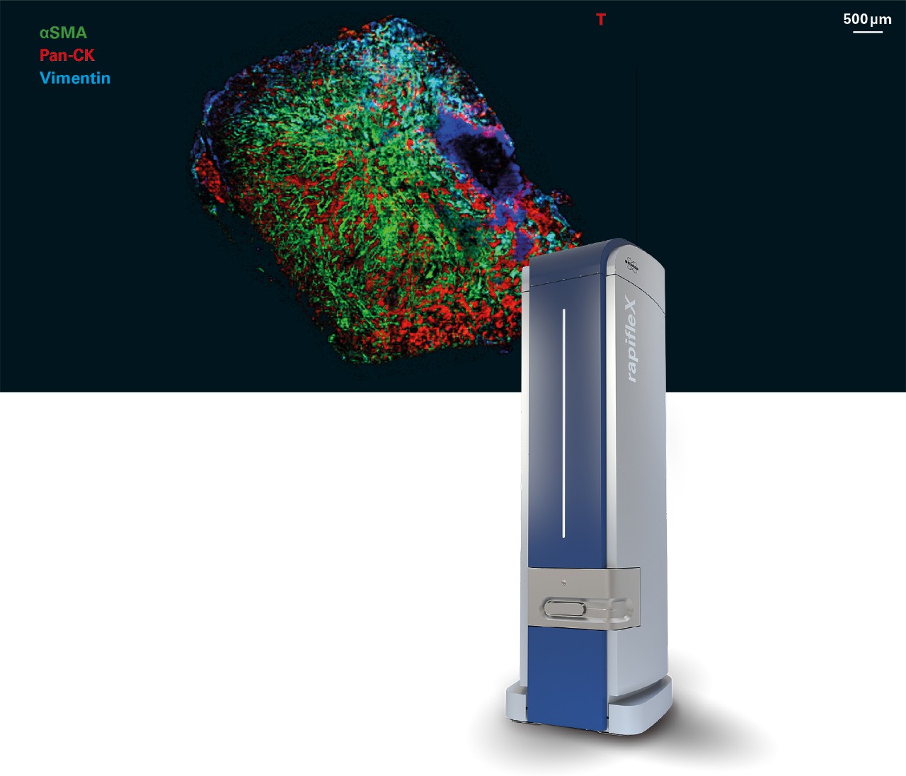
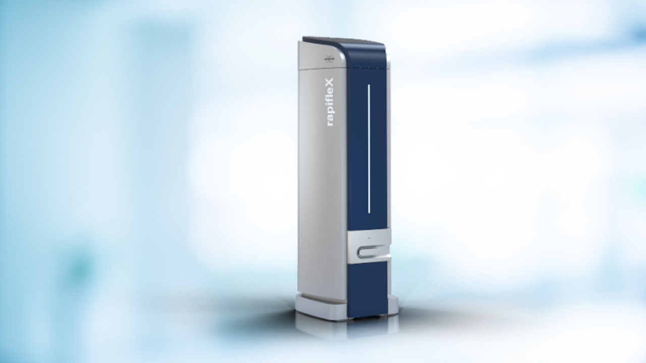
rapifleX® – the fastest MALDI Imaging platform available
- Quickly acquire targeted protein images from fresh frozen or FFPE tissue sections
- Maximize protein multiplex level without increasing analysis time
- Simple operation, easy to use and maintain with automated setup features for reproducible and consistent analysis
Bring Multiomics Intelligence to your disease-based research
Immuno-oncology: Visualize the Tumor Microenvironment
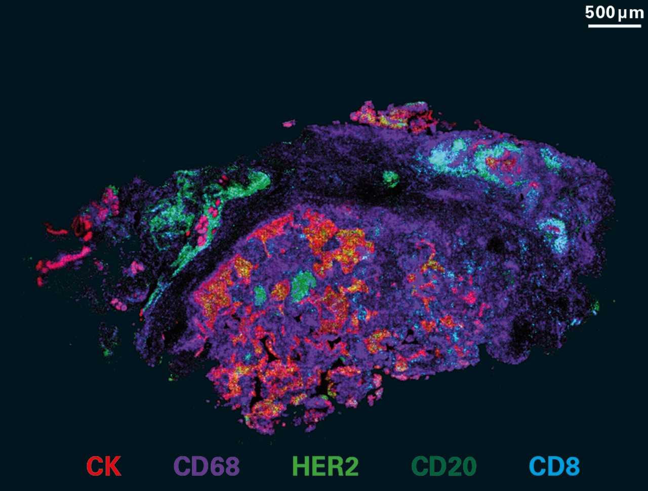
- Immuno-oncology focused markers for HiPLEX information gain
- Combine tumor microenvironment protein markers with metabolic functions in a multiomic overlay
- Accurately assess immune response, determine protein activity, and cell signaling
- Tumor subtyping realized with software solutions from SCiLS™ Lab to aid in classification
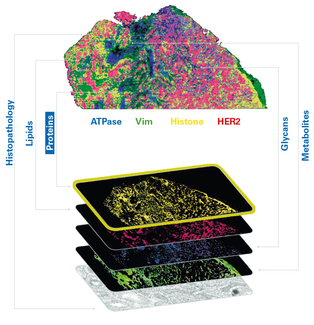
Expand functionality into glycobiology
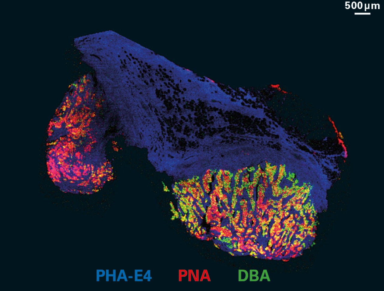
- Enable multiomics capabilities with glycans to uncover cellular signaling pathways
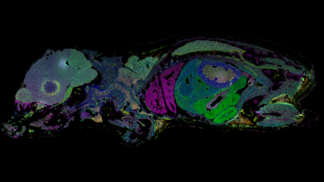
- Breast cancer images can reveal aberrant glycosylation (shown) and co-localization with protein markers
Neural Degeneration Profiling
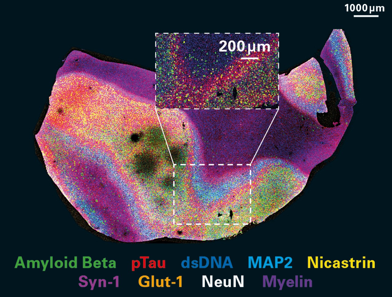
- Neurodegenerative disease visualization for key protein differentiators and structural disease pathology
- Human Alzheimer’s brain shown with amyloid plaques (Aβ) and neurofibrillary tangles (pTau)
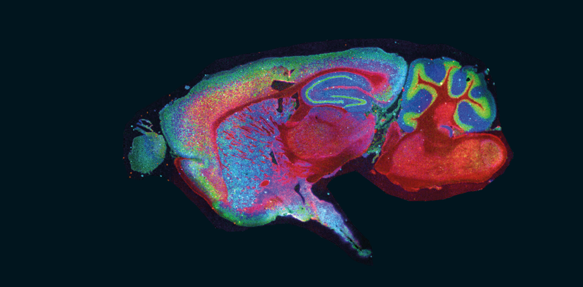
Neuroscience: Mapping Molecular Structure for Translation
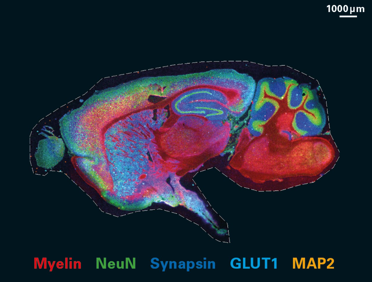
- Neurological overview of neurons and glial cells for multiple molecular functions
- Assess the structural and spatial organization of neurobiological areas of interest
Utilize our expertise to reduce your workload with advanced software for automation, high-quality consumables for reproducibility, and continued training and support

For Research Use Only. Not for use in clinical diagnostic procedures.