Bioinorganic Chemistry
Bioinorganic Chemistry and its role in modern chemistry
Right in the middle, between biology, organic, and inorganic chemistry, bioinorganic chemistry has found its place as an important contributor in the research of metal-based, biologically relevant complexes and enzymes. The highly interdisciplinary work includes the design, synthesis and characterization of model systems and extends into biomineralization and solid-state chemistry.
The investigation of biochemical processes in the various states of aggregation on a molecular or even atomic level requires analytical instrumentation providing the highly accurate information in the desired time domain. Our long-standing strong partnership with leading experts in the field enables us to offer the instrumentation essential for getting answers to the challenges in today’s bioinorganic chemistry.
Vibrational Spectroscopy in Bioinorganic chemistry
FT-IR and Raman analysis easily bridge the gap between inorganic and biological materials and provide full characterization of both.
Analysis of gas-processing metalloenzymes
Iron, nickel, copper, molybdenum and vanadium are essential components of cellular gas metabolism in all aspects of life. In association with biological macromolecules, gas-processing metalloenzymes (GPMs) are formed to catalyze a variety of redox reactions. In this example, FT-IR spectroscopy was introduced for the analysis of GPMs such as cytochrome c oxidase, nitrogenase and hydrogenase. Infrared spectroscopy provided information on the geometry and redox state of catalytic cofactors, protonation state of amino acid residues, hydrogen bonding network and structural changes of the protein.
Development of dental implants
Teeth are a high-performance bioinorganic product and filling cavities must at the least happen with adequate material. Raman microspectroscopy can help in the development of new filling materials. In this example, Raman microscopy was used to identify the homogeneity of a dental composite made of Dimethacrylat and glass/ceramics.
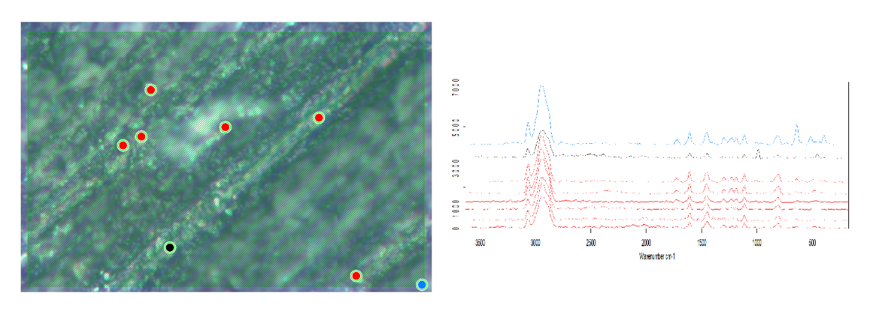
Left: Dental filling material and Raman measurement points.
Right: Raman spectra of polymer matrix and additives.
Study of skeletal remains
Bone is studied by IR macroscopically and also by IR Imaging for biopharmaceutical/medical and archeological purposes.This is used to clarify the origin of bone material, as well as to determine the mineralization degree and to investigate the aging process of the specimens. In this example the bone mineral was tracked in several forensic and archeological samples of skeletal remains.
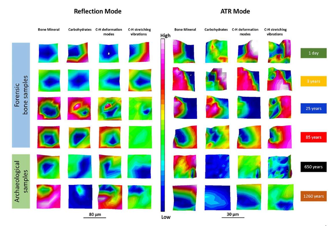
Comparison of FT-IR images of forensic and archeological bone samples.
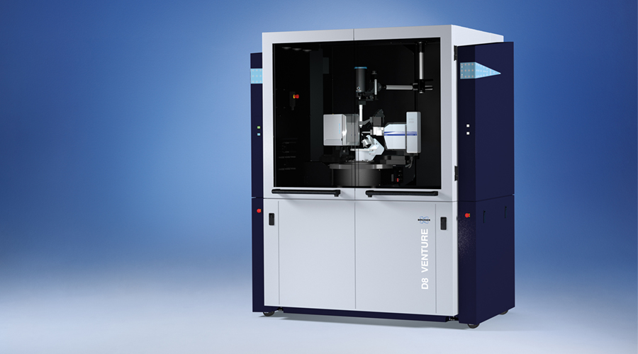
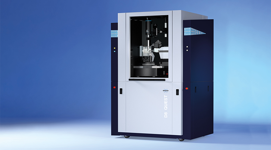
Single Crystal X-ray Diffraction in Bioanorganic Chemistry
Modern bioinorganic chemistry on metal-containing proteins, enzymes, and other biomolecules largely benefits from the capability of single crystal X-ray diffraction (SC-XRD) to determine the three-dimensional structure of a molecule or complex in atomic detail. Often with limited effort a deep understanding of the mechanisms driving the biological processes involving metal ions can be obtained.
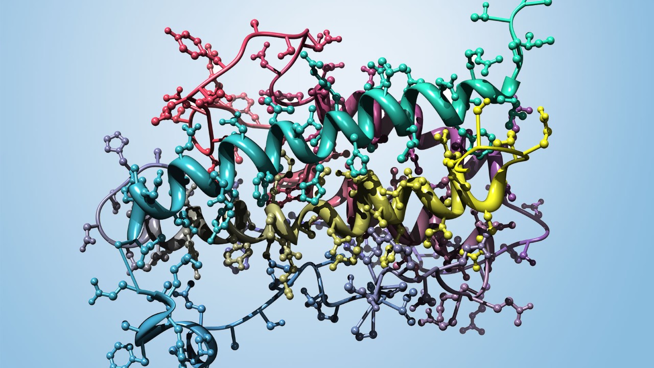
Literature Examples of XRD use in bioinorganic chemistry
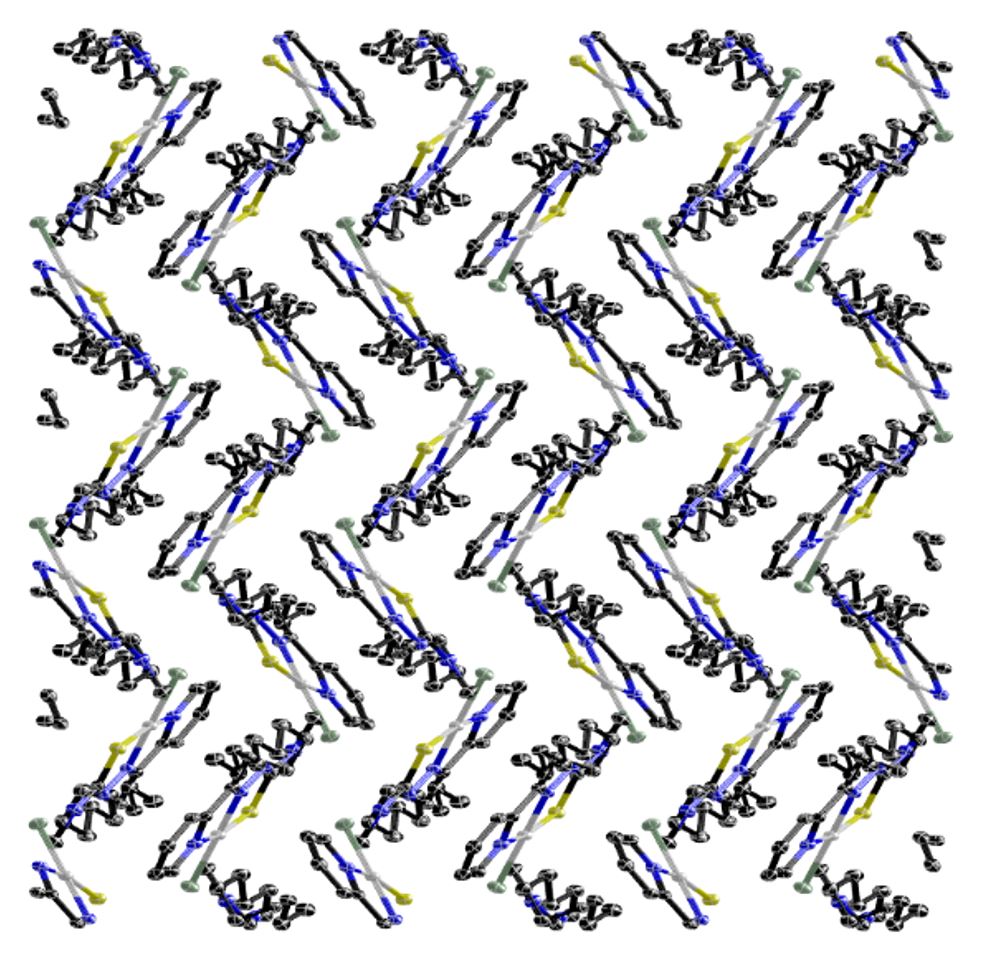
Platinum(II)c complexes are playing an important role as anticancer agents. Newly synthesized Pd(II) and Pt(II) thiosemicarbazone complexes were studied on four human glioblastoma cell lines. An n-decyl derivative has been investigated in detail using X-ray crystallography and coordination geometry around the metal atom has been determined. Next to Cl···H-N bond the long aliphatic side chain seems to have significant influence on the arrangement of the molecules in the crystal, leading to a “stair-case” arrangement of the individual molecules and the long alkyl chains alternatingly pointing outwards.
Guanine quadruplexes are potential candidates in anticancer therapeutics. This study reports a systematic investigation of a family of luminescent platinum(II) complexes devised to overcome topological challenges in the field by adding an additional recognition site and various linker moieties. Single crystals X-ray diffraction analysis reveals a significant influence of the linker length on the overall three-dimensional appearance and helps to draw first conclusions with respect to the potential of this new approach.