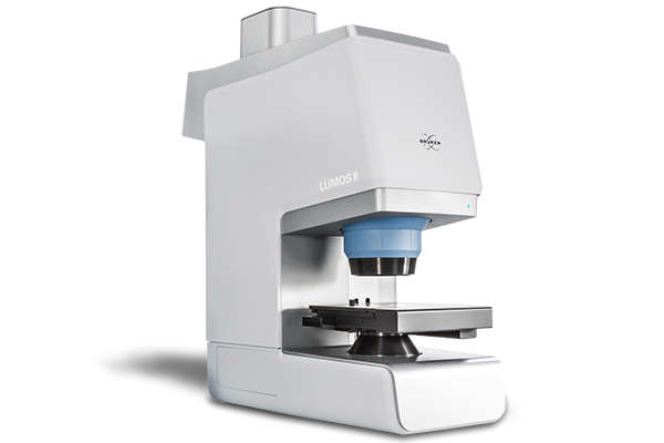
LUMOS II
Exceptional IR.
Brilliant Visuals. Ultrafast Imaging.
Infrared Microscopy the way it was meant to be.
More space for sample preparation. More speed for chemical imaging. More perfomance in ATR, transmission and reflection microscopy. This is what we call a true game changer - no discussion.
Single-point FT-IR microscopy? FPA imaging? IR laser? Your choice.

FT-IR microscopy:
- TE-MCT as standard detector
- Industry leading ATR crystal design
- No liquid nitrogen, no dry-air purge
- LN-MCT and DTGS optionally available
- Up to two single-element detectors in parallel
- Upgrade to FPA imaging anytime
- Analyze samples of up to 40 mm in height

FT-IR FPA imaging:
- Focal-plane array (FPA) imaging detector
- Create FT-IR images at 1.6 mm² per sec
- Access 1.25 µm spatial resolution (ATR)
- FT-IR images at full MIR spectral range
- Imaging in ATR/TRANS/REFL
- Patented PermaSure+ imaging calibration
- Up to two additional detectors in parallel

IR laser imaging (ILIM):
- Room temperature focal-plane array detector
- Create IR images at 4.5 mm² per second
- IR laser images at 1,800 - 950 cm-1
- Create chemical images in TRANS and REFL
- Patented spatial coherence reduction
- Application workflows (Tissue, Particle, Tablet)
- Open design laser class 1 instrument
No matter what you choose, LUMOS II always offers ...
Easy to use all in one software.
The LUMOS II Wizard.

Inertness against high humidity.
Using inert optical materials.

AI powered evaluation routines.
Adaptive Chemical Imaging.

Compliance to cGMP and FDA 21 CFR p11.
Comprehensive audit trail.

Complete automization and long service-life.
Automated Knife-Edge Apertures.

Open design with easy access to samples.
270° access from three sides.

The FT-IR Microscopy and IR Imaging Powerhouse
The Anywhere / Anyone FTIR Microscope

We believe that it is high time to make advanced techniques available to every user, regardless of their skill level. The benefits of FT-IR imaging and microscopy are too great to restrict access by cumbersome hard- and software.
From the start the LUMOS II was meant to make FT-IR imaging faster, easier, more accurate and reliable – and even more fun. Of course, this required us to include new and improve upon proven technology.
That's why we tailored the LUMOS II, its software and user interface specifically to the user. Beginners get perfect results in no time, while experts maintain total instrument control.
Superior µ-ATR FT-IR Capabilities

It comes down to this: Better instrument. Better results.
Whether it is transmission, reflection or attenuated total reflectance (ATR), the LUMOS II is always the right choice. But its greatest strength is ATR microscopy enhanced by FPA technology. This makes the LUMOS II a universal tool for failure analysis and product development.
To cut a long story short, its ATR capabilities are unsurpassed. Period. Don‘t settle for unreliable, manual ATR accessories – get the best. Get the LUMOS.
The retractable crystal is controlled by high precision piezoelectrical motors and integrated into the lens. This allows you to enjoy a clear view of the sample while your measurement still takes place exactly where you want it.
Innovation in FT-IR Microscopy in Every Fiber

Endurance and power for your applications.
For us it is a natural thing to pass on the best technology to our customers. Of course, this also applies to the LUMOS II.
The RockSolidTM interferometer guarantees constant performance, while modern electronics ensure mechanical precision and low energy consumption. Meanwhile, the software monitors instrument effectiveness and always ensures correct functionality.
LUMOS II Application Videos
Choose your µ-FT-IR Application
Particle & Surface Analysis

Industrial Manufacturing

Environmental Science

Life Science

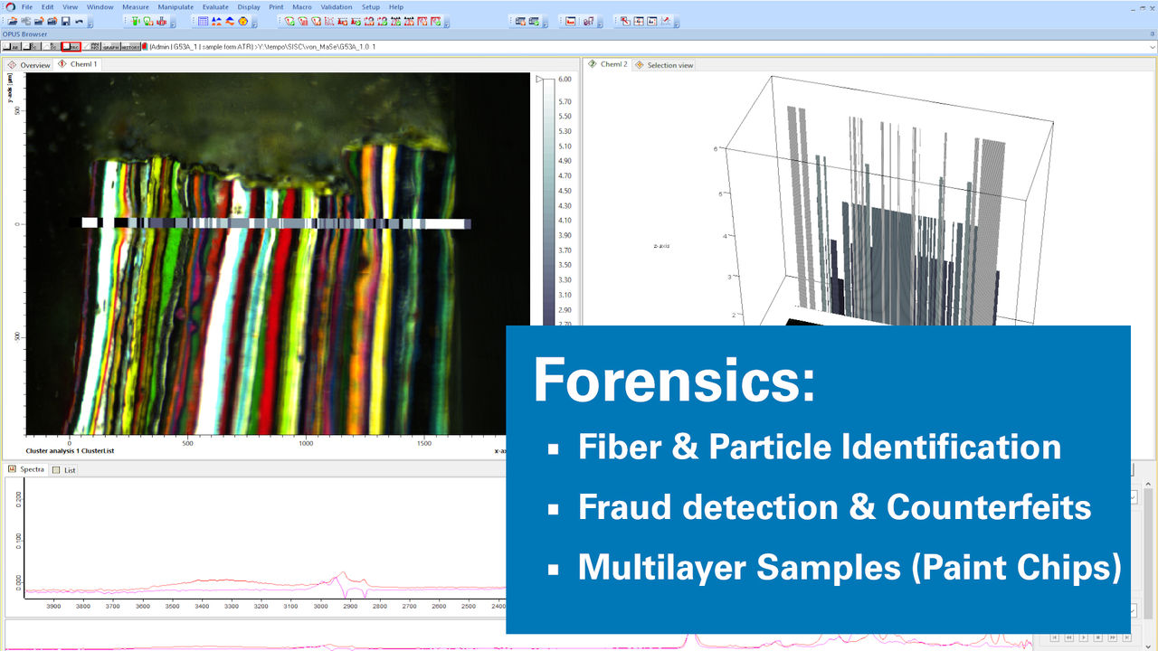
Forensics
Polymers and Plastics

Art and Restoration

Pharmaceuticals

Surface Science


Microscopy Workshop Eindhoven: Imaging
Pharmaceutical Manufacturing: Eyetec Belgium
OPUS Release 9.0 Highlights for LUMOS II | Q1 2025
New Feature: The Autonomous Composition Identifier (A.I.D.) simplifies search and identification.
The Autonomous Composition Identifier (A.I.D.) is an AI-driven software tool for identifying the chemical composition of samples based on IR or Raman spectral data. It incorporates the previous O/SEARCH features and adds a multi-step algorithm to find the best match from reference spectral databases, whether dealing with pure substances or complex mixtures. A.I.D. intelligently evaluates and recommends optimal matches with a balance of reliability and sensitivity—automatically generating diagnostics and visual reports, including residual spectra and hit quality graphs. It delivers high-performance results in seconds and supports in-depth, interactive analysis of alternative identifications.

New Feature: Co-local Measurement – Unified Analysis Across IR and Raman
The Co-local Measurement function, part of the OPUS/OBJECT package, enables seamless analysis of the same region of interest on microscopic samples using both IR and Raman microscopy. It allows the transfer of overview images and measurement areas between LUMOS II, HYPERION II, and SENTERRA II instruments. This ensures that IR and Raman data are collected from precisely the same spot, enabling complementary chemical insights. Co-local Measurement bridges the gap between two techniques, improving data correlation and boosting confidence in results for heterogeneous or complex samples.

Updated Feature: OPUS/OBJECT now combines "Find Particle" and "Cluster ID"
The Particle Identification function (part of the OPUS/OBJECT package) combines the power of “Find Particles” and “Cluster ID” into one streamlined workflow. It detects particles within IR and Raman chemical images, determines their dimensions, and identifies their chemical makeup—all in one go. This integrated approach simplifies analysis and accelerates decision-making for complex samples.

OPUS Release 8.8 Highlights for LUMOS II | Q3 2023
New Feature: 3D FocusFusion now allows the creation of visual images of infinite "sharpness"
This new feature creates a pin sharp visual image, even if the sample has a rough surface or isn’t flat at all.
This infinite "sharpness" in microscopic images helps in region of interest selection. Now it will be possible to generate sharp visual images of samples for which the depth of field of the LUMOS II is limited.

New Feature: FPA imaging can now follow round / circular measurement areas
Now, the measurement grid can be placed in a round shaped way to measure a whole particle filter with FPA imaging. By only measuring the actual region of interest users safe significant amounts of time during FPA measurements of round regions of interest like filters in microplastic analysis.

OPUS Release 8.7 Highlights for LUMOS II | Q3 2021
New Feature: High Performance Chemical Image Generation by New Adaptive K-means Clustering Function
This new function is the logical next development step for our well known Cluster analysis function.The Adaptive K-means Clustering Function is based on a new algorithm, which enables a non-supervised and autonomous determination of spectral variance within your imaging or mapping results.
- Forecasting or time consuming searching of the included amount of chemical classes is no longer been necessary as the algorithm can predict all included chemical classes by itself.
- This major function is important for all kinds of chemical imaging and distribution analysis of unknown samples or small structures within larger datasets.
- Together with the LUMOS II analysis and evaluation is as easy as possible and safes your valuable time and nerves.
New Feature: “Cluster ID” Function for Identification of Classes in 3D Spectral Data
Our new Cluster ID function enables the identification of clusters within imaging and mapping data using the OPUS functions: spectrum search in libraries, quick compare, or identity test.
- Easy determination of the chemical identity of classified sample components for particles, layers in laminates, components of pharmaceutical tablets and other inhomogeneous materials.
- Reliable and comprehensive statistics reports about quantity, size and of course identity of all analyzed structures is provided and leads particle and technical cleanliness analysis to a new, autonomous level.
Updated Feature: "Find Particles" function now contains a novel particle detection method
The proven "Find Particle" software can now be applied to both: the visual and the IR image. With this updated feature, you are able to do particle detection based on chemical images that were measured by the LUMOS II.
- While particle recognition for low contrast structures and off-white/transparent particles/fibers on off-white filter membranes can be tedious, a postrun particle determination based on the chemical IR image allows you to determine quantity and size of particles from your imaging or mapping results.
- With the Find Particle Function together with the LUMOS II you will never miss any detail – neither in visual nor in IR range.


FT-IR Microscopes Literature Room
Learn more about our FT-IR microscopes and solutions by downloading related literature.


