Novel Applications of Optical Tweezers in the Physical and Life Sciences
Simultaneously manipulate molecules and nanoparticles and explore the piconewton forces acting on them.
In this mini-symposium, our special guest-experts discuss the role OT can play in the search for new and innovative analysis and characterization methods, and how the technique can contribute towards a more in-depth understanding of nanomechanical processes.
Understand The Role OT Plays in Innovation and Discovery
The Nobel Prize winning Optical Tweezers (OT) technique enables the non-invasive, nanometer-precise manipulation of molecules and nanoparticles and the simultaneous application and recording of the piconewton forces acting on them. OT can be used to perform optical, thermodynamic or nanomanipulation experiments, to trigger, visualize, and quantify molecular interactions, and to characterize mechanical properties such as adhesion, viscoelasticity, or deformation.
Presentations in this symposium include an introduction to Novel Applications of Optical Tweezers in the Physical and Life Sciences, as well as individual sessions focused on how OT can be used to study:
- The ageing behavior of colloidal suspensions;
- DNA repair mechanisms; and
- The interactions between immune receptors and the cytoskeleton in T cell mechanotransduction.
Find out more about our optical tweezers technology:
Presentation Abstracts
Contact and Macroscopic Ageing in Colloidal Suspensions
Dr. Julie Goyon and Antoine Aubel, Laboratoire Navier, Université Gustave Eiffel - ENPC - CNRS, France
While the ageing behavior of dense suspensions or pastes at rest is almost exclusively attributed to structural dynamics, a set of experiments conducted on dense aqueous silica suspensions allowed us to identify another ageing process: contact-controlled ageing. We have investigated the origin of shear modulus and yield strength ageing in dense aqueous silica suspensions at moderate ionic forces. By combining rheometry and confocal microscopy, we first showed that the elastic modulus and yield stress of these suspensions at rest grow logarithmically with time, while their structural evolution is rapidly arrested by the formation of interparticle contacts [[i]]. So these suspensions age in the absence of structural rearrangement. Then, by performing three-point optical tweezer (OT) bending tests on particle rods, we showed that both the rolling stiffness and rolling threshold grow logarithmically in time. By comparing the results of the measurements on the particle contact properties with the results of the rheometry tests, we were able to show that ageing of interparticle contacts governs both shear modulus ageing and yield stress ageing of these dense aqueous silica suspensions [[ii]-[iii]].
REFERENCES:
- i Fusier, J. et al. (2018). Rheology signature of flocculated silica suspensions. Journal of Rheology, 62(3), 753-771
- ii Bonacci, F. et al. (2020). Contact and macroscopic ageing in colloidal suspensions. Nature Materials, 19(7), 775-780
- iii Bonacci, F. et al. (2021). Yield Stress Aging in Attractive Colloidal Suspensions. Phys. Rev. Lett. 128, 018003
Probing the Biomechanics of the Interaction between RuvA and DNA Holliday Junctions
Dr. Soma Dhakal, Virginia Commonwealth University, USA
DNA Double-strand breaks (DSBs) are among the most detrimental types of DNA damage. The repair of DSBs occurs via a process called homologous recombination (HR), which involves several DNA-binding proteins. Therefore, to have a better insight into the repair mechanism and origin of repair defects, we need a better understanding of how these proteins interact with DNA and DNA intermediates such as Holliday junctions (HJs). Using dual-trap optical tweezers, we recently studied the biomechanics of HJ and a prokaryotic protein RuvA and showed that the RuvA protein not only stabilizes the HJ but also assists in its refolding.
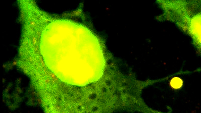
Probing Mechanical Interactions of Immune Receptors and the Cytoskeleton by Membrane Nanotube Extraction
Dr. Pierre-Henri Puech, INSERM, Adhesion & Inflammation, Marseille, France
In our labs, we use a collection of force based biophysical techniques to study early T cell mechanotransduction (Atomic Force Microscopy (AFM)1,2, Biomembrane Force Probe (BFP), Flow Chamber3, Traction Force Microscopy, Optical Tweezers), smart decorated substrates, and advanced quantitative surface microscopies4–6. We focus on the particular moment where the ligation of the T cell receptors (TCR) with antigens displayed on partner substrates (antigen-presenting cell - APC) is the very first step in triggering the T cells response. It has been demonstrated that this passes through the formation of the so called 'immune synapse' (IS) that involves additional molecules like integrins and CD45 7.
The mechanosensitive nature of the T-cell/APC and the mechanics of the TCR-complex is currently being studied extensively, but the question of the existence, if any, of a direct physical interaction between the TCR and the cytoskeleton is still not known8. Such an interaction could have a profound impact on the motion and recruitment of TCR at the interface between the T cell and the partner cell or substrate. In particular, it may explain complex behaviour, like biphasic response to substrate elasticity. The proposed mechanism crucially depends on this coupling9,10.
To explore this link, we used optical tweezers to pull membrane tubes from T cells using different antibodies as a handle, each targeting a given protein present at the IS, here, TCR/CD3, LFA1, CD45. We also pulled nanotubes after disrupting the actin cytoskeleton using Latrunculin. We then developed a theoretical mechanical model that retains the key biophysical properties of the coupled system, consisting of the T cell and the bound receptor/ligand pair on one hand and the force transducer on the other.
The model provides a fine description of the force curves and the fitting strategy we developed allowed us to extract information on cellular and molecular viscoelastic parameters. Our model also enables the estimation of key viscoelastic parameters, which account for the stiffness of the ligand/receptor complex, the cell rigidity, and viscosity. To process our large datasets, composed of both membrane tube pulling and handle/protein detachment events, we developed a refined post-fit data processing strategy to recover molecular information from the latter in the frame of our model to strengthen our statistics.
Our results suggest that the mechanical link with the cytoskeleton is modulated for the different molecules in the immune synapse such as TCR, LFA-1 and CD45. Finally, our model allows us to propose a quantitative description of the intra-cellular molecular bonds, which are usually quite difficult to infer or impossible to measure, from externally pulling nano-tubes, a rather easy experimental method adaptable to other force-based techniques such as AFM or BFP.
REFERENCES:
- Sadoun, A. et al. Sci. Rep. 11, 6783 (2021).
- Zak, A. et al., Biophys. J. 120, 1692–1704 (2021).
- Limozin, L. et al., Proc. Natl. Acad. Sci. U. S. A. 116, 16943–16948 (2019).
- Sadoun, A. et al. Wiley Analytical Science https://analyticalscience.wiley.com/do/10.1002/was.00010013 (2020).
- Benard, E. et al., Front. Immunol., 9, 1–12 (2018).
- Dejardin, M.-J. et al., Nano Lett. (2018) doi:10.1021/acs.nanolett.8b03134.
- Limozin, L. et al., J. Membr. Biol. 252, 397–412 (2019).
- Puech, P.-H. et al., Open Biol. 11, 210256 (2021).
- Dillard, P. et al., Biophys. J. 107, 2629–2638 (2014).
- Wahl, A. et al. Proc. Natl. Acad. Sci. 116, 5908–5913 (2019).
Speakers
Dr. Julie Goyon
Laboratoire Navier, Université Gustave Eiffel - ENPC - CNRS, FranceDr. Julie Goyon’s research interests are in the field of physics, the physical chemistry of materials, and their applications, in particular in civil engineering materials. In her work, she aims to establish the link between mechanical properties (macroscopic) and the local physical or physico-chemical properties (microscopic) of materials in order to predict macroscopic properties and to understand their physical and/or physico-chemical origin. During the last few years, she has been particularly interested in the link between formulation/microstructure and material properties in model materials such as dense silica suspensions.

Antoine Aubel
Laboratoire Navier, Université Gustave Eiffel - ENPC - CNRS, FranceAntoine Aubel graduated from ENSCI Limoges. His Ph.D. work at Navier Lab with Xavier Chateau and Julie Goyon focuses on rheology of model cement pastes at an early age. The goal of this study is to better understand how chemical additives such as superplasticizers modify colloidal interactions, which control the behavior of fresh cement pastes. By combining rheometry tests and experiments with laser tweezers, they try to link the rheological properties of these pastes to the contact properties between particles. This fundamental understanding can help to better control the formulation of cementitious pastes used in industrial processes.

Dr. Soma Dhakal
Virginia Commonwealth University, USADr. Soma Dhakal received his Ph.D. from Kent State University, USA, where he worked with Prof. Hanbin Mao on the mechanical unfolding of DNA secondary structures (i-motifs and G-quadruplexes) using optical-tweezers. In his postdoctoral research with Prof. Nils G. Walter at the University of Michigan, he applied single-molecule fluorescence microscopy in nanotechnology applications, which involved the actuation and monitoring of single enzyme activities. Currently, he is an assistant professor in the department of chemistry at Virginia Commonwealth University (VCU), USA, where he studies protein/DNA biophysics using fluorescence microscopy and optical tweezers and develops FRET-based single-molecule sensors. His complete list of publications can be found on the Google scholar page:
(https://scholar.google.com/citations?user=2gj2R78AAAAJ&hl=en).

Dr. Pierre-Henri Puech
INSERM, Adhesion & Inflammation, Marseille, FranceDr. PH Puech was trained as a soft condensed matter physicist (Ph.D. with F. Brochard, Institut Curie, Paris, in collaboration with PG de Gennes) and gradually turned towards biology (Postdoc in Max Planck Institute for Cell Biology and Genetics & TU Dresden with D. Müller & CP Heisenberg). He currently works as a senior researcher for INSERM in the Lab Adhesion and Inflammation in Marseille, France, with an emphasis on T cell mechano-transduction. He is a specialist in force based techniques: AFM (and pioneer in SCFS), micropipettes and optical tweezers. He is part of Institut Convergence Centuri (Marseille) and several French research consortiums (GDRs). He chaired the EMBO WS / Les Houches School of Physics on Immunobiophysics (2021), organizes a virtual immuno-biophysics seminar, and will chair the 2023 edition of this conference (immunobiophysics.org).