Bruker Online Neuroscience Summit 2026:
Innovations in Brain Research
Bruker Online Neuroscience Summit 2026:
Innovations in Brain Research
This summit will showcase groundbreaking research from leading researchers utilizing advanced imaging technologies, including Bruker two-photon microscopes, single-photon miniscopes, and super-resolution microscopes, to explore neuronal activity in the brain. Don't miss this chance to connect with the scientific community and stay at the forefront of neuroscience innovation.
Abstracts and Speaker Bios
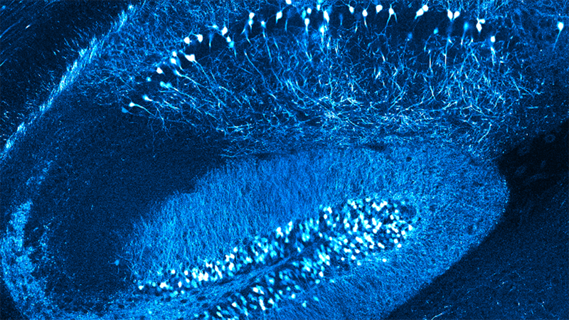
Abstract: Using state-of-the-art high-resolution two-photon imaging combined with chemogenetic and optogenetic circuit manipulations, head-fixed behavioral paradigms for spatial and contextual learning, and analytical approaches including automated ROI segmentation and ML-based image processing approaches, we are studying behaviorally driven compartment-specific dynamics in synapses, axons, dendrites, soma, cell types, and ensembles in the cortico-hippocampal circuit. First, we found that the activity dynamics and temporal stability of spatial representations in soma versus dendrites of hippocampal pyramidal neurons differ (Moore et al., Nature Communications, 2025) and may be modulated by GABAergic microcircuit dynamics. Finally, we found that entorhinal cortex axonal projections to the hippocampus convey both contextually salient and spatially tuned input and are modulated by hippocampal feedback projections to the entorhinal cortex (Butola et al., Nature Neuroscience, 2025). Glutamatergic and GABAergic input from both LEC to CA1 (Basu et al . Science 2016; Bilash et al., Cell Reports 2023, Hernandez Frausto in preparation) versus CA3 (Robert et al., Science 2025) targets different interneuron microcircuits to differentially modulate excitatory output in a compartment and pathway-specific manner and play distinct functional roles in learning vs. recall of memory behaviors. The Glutamatergic and GABAergic LEC inputs conjunctively modulate learning and context-driven stability and remapping of place maps in CA3 (Robert et al., Science 2025), whereas glutamatergic input from LEC is crucial for supporting memory recall and stability of task-selective place ensembles during a high-demand goal-oriented learning task (Hopkins et al., 2026 bioRxiv; Zemla et al., Cell Reports, 2022).

Jayeeta Basu, Ph.D.
Associate Professor, Department of Neuroscience, Department of Psychiatry
WEBSITE:
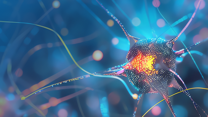
Abstract: Activation of voltage-gated calcium channels at presynaptic terminals leads to local increases in calcium and the fusion of synaptic vesicles containing neurotransmitter. Presynaptic output is a function of the density of calcium channels, the dynamic properties of the channel, the distance to docked vesicles, and the release probability at the docking site. We demonstrate that at Caenorhabditis elegans neuromuscular junctions two different classes of voltage-gated calcium channels, CaV2 and CaV1, mediate the release of distinct pools of synaptic vesicles. CaV2 channels are concentrated in densely packed clusters ~250 nm in diameter with the active zone proteins Neurexin, α-Liprin, SYDE, ELKS/CAST, RIM-BP, α-Catulin, and MAGI1. CaV2 channels are colocalized with the priming protein UNC-13L and mediate the fusion of vesicles docked within 33 nm of the dense projection. CaV2 activity is amplified by ryanodine receptor release of calcium from internal stores, triggering fusion up to 165 nm from the dense projection. By contrast, CaV1 channels are dispersed in the synaptic varicosity, and are colocalized with UNC-13S. CaV1 and ryanodine receptors are separated by just 40 nm, and vesicle fusion mediated by CaV1 is completely dependent on the ryanodine receptor. Distinct synaptic vesicle pools, released by different calcium channels, could be used to tune the speed, voltage-dependence, and quantal content of neurotransmitter release.

Brian D. Mueller, Ph.D.
Postdoctoral Researcher,
Charité – Universitätsmedizin Berlin, University of Utah
WEBSITE:
Abstract: Sensory perception is shaped by experience, giving stimuli behavioral significance. Basal forebrain (BF) cholinergic neurons in mice, which are crucial for arousal and motivation, also regulate sensory processing. Within BF nuclei, glutamatergic (vGlut2BF) neurons receive cholinergic input and modulate behaviors, but their roles in encoding sensory significance remain unclear. Using in vivo calcium imaging, we found that vGlut2BF neurons initially poorly encoded odor identity. However, their response to conditioned odors increased following associative learning, and their population activity more distinctly encoded paired stimuli, reflecting emergent value representation. Furthermore, pairing stimulation or inhibition of vGlut2BF neurons with specific odors altered odor preferences, suggesting that appropriately timed vGlut2BF neuronal activity is sufficient to influence valence assignment. Our findings reveal that vGlut2BF neurons transform sensory input into motivationally significant stimuli, positioning the BF as a key hub for linking sensory processing with motivational states and experience-driven plasticity.

Abstract: Understanding how individual neurons contribute to specific behaviors requires precise neural activity imaging aligned with well-designed behavioral paradigms. This talk presents a framework for investigating behavior-time-locked neuronal roles using single-cell-level neural activity imaging in freely moving mice. We emphasize the importance of designing behavioral tasks that directly test neuroscience hypotheses, ensuring precise temporal alignment between neural activity and behavioral events. Understanding how individual neurons contribute to specific behavioral processes, such as food discovery, approach, consumption, reward learning, and extinction memory, requires precise neural activity imaging aligned with well-structured behavioral paradigms. We emphasize the importance of designing behavioral tasks that capture key moments in reward-related behaviors, ensuring precise temporal alignment between neural activity and behavioral transitions. By integrating advanced single-cell-level neural activity imaging mini1P and mini2P techniques with hypothesis-driven behavioral paradigms, we aim to elucidate the neuronal mechanisms underlying reward processing and learning, providing new insights into neural circuit function.

Abstract: Using cellular-level two-photon optogenetic perturbations in ferret visual cortex, we show that excitatory neurons exert spatially broad suppression onto neighboring cells — contrary to classic Mexican-hat predictions. This suppression is contrast-dependent: functionally coupled, similarly tuned cell pairs switch from facilitation under low contrast to strong suppression at high contrast. Network modeling identifies a "Cross-Dominant" connectivity regime — where inhibitory-to-excitatory coupling dominates — as the mechanism underlying this reversal, confirmed by inhibitory cell-type-specific perturbations. To extend these findings to the population level, we are implementing 3D holographic photostimulation to simultaneously target spatially distributed ensembles defined by columnar identity and functional tuning, directly testing whether Cross-Dominant dynamics govern multi-cell ensemble interactions during flexible sensory processing.

Benjamin Scholl, Ph.D.
Assistant Professor, Dept. of Physiology and Biophysics, University of Colorado School of Medicine
WEBSITE:
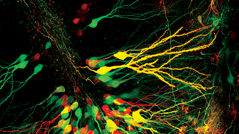
Abstract: Neurological tissue is organized across a wide range of scales from tissue down to synapses and sub-synaptic structures. To derive a complete understanding of neuroscience, we must come to understand the function and organization of the synapse. Synapses have historically been difficult to study directly due to their very small size (on the order of ~100nm). While Single Molecule Localization Microscopy (SMLM) offers high enough resolution for sub-synaptic studies(20nm resolution lateral, or about 1/5th of a synapse), traditional SMLM instruments aren't compatible with many neurological preps, as isolated neurons, tissue sections and whole mount model organisms are far too large. The Vutara VXL SMLM microscope has been designed to handle just these specimens, allowing for imaging at sub-synaptic resolution across a wide array of neuroscience samples and models.
In this talk, I'll cover a wide variety of applications from neurons in culture, sections of tissue from a neurodegeneration model, to imaging living neurons in culture and C. elegans, with a focus on the synapse and its structure; demonstrating the versatility of the Vutara VXL as a tool for high resolution neurological discovery.

Abraham Kohrman, Ph.D.
Applications Specialist for Biological Microscopy, Bruker FM
WEBSITE: