|
2024
|
bioRxiv
|
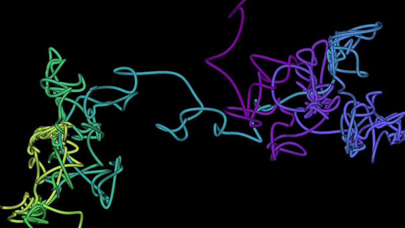
The molecular mechanism of on-demand sterol biosynthesis at organelle contact sites.
|
Zung N, Aravindan N ... Schuldiner M
|
|
|
2024
|
Bioprotocol
|
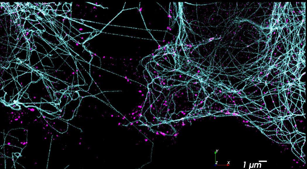
Using Localization Microscopy to Quantify Calcium Channels at Presynaptic Boutons.
|
Brian D. Mueller, Sean A. Merrill ... Erik M. Jorgensen
|
Direct Imaging of Synaptic Structure
|
|
2024
|
Nature Communications
|
Functional specificity of liquid-liquid phase separation at the synapse.
|
Natalie J. Guzikowski and Ege T. Kavalali
|
Synaptic Organization, Control of Signalling
|
|
2024
|
Science
|
Native architecture of a human GBP1 defense complex for cell- autonomous immunity to infection.
|
Zhu S, Bradfield CJ ... MacMicking JD
|
Cells; Membrane; Bacteria; Cellular Structures
|
|
2024
|
bioRxiv
|
GABA and astrocytic cholesterol determine the lipid environment of
GABA(A)R in cultured cortical neurons.
|
Yuan Z, Pavel MA ... Hansen SB
|
|
|
2024
|
Nano Lett
|
One Stone, Two Birds: High-Brightness Aggregation-Induced Emission Photosensitizers for Super-Resolution Imaging and Photodynamic Therapy.
|
Wang Z, Zhou Y ... Meng L
|
Diseases; Eukaryota; Chemicals and Drugs
|
|
2024
|
Microsc Microanal
|
Indirect Correlative Light and Electron Microscopy (iCLEM): A Novel
Pipeline for Multiscale Quantification of Structure From Molecules to Organs.
|
Struckman HL, Moise N ... Veeraraghavan R
|
Cells; Membrane; Cellular
Structures; Microscopy
|
|
2024
|
bioRxiv
|
Increased interaction between connexin43 and microtubules is critical for glioblastoma stem-like cell maintenance and tumorigenicity.
|
Smyth JW, Guo S ... Lamouille S
|
|
|
2024
|
J Am Chem Soc
|
Targeted Photoconvertible BODIPYs Based on Directed
Photooxidation-Induced Conversion for Applications in Photoconversion and Live Super-Resolution Imaging.
|
Saladin L, Breton V ... Collot M
|
Cells; Nervous System; Eukaryota;
Chemicals and Drugs
|
|
2024
|
Nature
|
RNA-mediated symmetry breaking enables singular olfactory
receptor choice.
|
Pourmorady AD, Bashkirova EV ... Lomvardas S
|
Cells; Nervous System; Genetic
Phenomena; Genetic Structures
|
|
2024
|
Elife
|
Mechanical activation of TWIK-related potassium channel by
nanoscopic movement and rapid second messenger signaling.
|
Petersen EN, Pavel MA ... Hansen SB
|
Cells; Membrane; Signal
Transduction; Cellular Structures
|
|
2024
|
bioRxiv
|
Enhancer-promoter hubs organize transcriptional networks
promoting oncogenesis and drug resistance.
|
Perlman BS, Burget N ... Faryabi RB
|
|
|
2024
|
bioRxiv
|
Long axial-range double-helix point spread functions for 3D
volumetric super-resolution imaging.
|
Nakatani Y, Gaumer S, Shechtman Y, and Gustavsson AK
|
|
|
2024
|
J Extracell Vesicles
|
Characterisation of LPS+ bacterial extracellular vesicles along the gut-
hepatic portal vein-liver axis.
|
Jain H, Kumar A ... Deep G
|
Cells; EV; Bacteria; Cellular
Structures
|
|
2024
|
bioRxiv
|
Lysine Demethylase 4A is a Centrosome Associated Protein Required
for Centrosome Integrity and Genomic Stability.
|
Chowdhury P, Wang X ... Dere R
|
|
|
2024
|
Neurophotonics
|
Molecular mapping of neuronal architecture using STORM microscopy and new fluorescent probes for SMLM imaging.
|
Breton V, Nazac P ... Danglot L
|
|
|
2024
|
ACS Omega
|
Fast In Vitro Synthesis and Direct Labeling of Nanobodies for Prototyping in Microscopy Applications.
|
Behrens L, Walter RM ... Zemella A
|
|
|
2024
|
Methods Mol Biol
|
Quantitative Super-Resolution Imaging of ER-Phagy Initiation in Cells.
|
Balakrishnan A, Glogger M ... Heilemann M
|
Cells; Organelles; Cellular Structures;
Microscopy
|
|
2023
|
Nat Cell Biol
|
Curved adhesions mediate cell attachment to soft matrix fibres in
three dimensions.
|
Zhang W, Lu CH ... Cui B
|
Cells; Membrane; Cellular
Structures; Chemicals and Drugs
|
|
2023
|
bioRxiv
|
Curved adhesions mediate cell attachment to soft matrix fibres in 3D.
|
Zhang W, Lu CH ... Cui B
|
|
|
2023
|
Membranes (Basel)
|
Cholesterol Regulation of Membrane Proteins Revealed by Two-
Color Super-Resolution Imaging.
|
Yuan Z, and Hansen SB
|
|
|
2023
|
Nature
|
PLSCR1 is a cell-autonomous defence factor against SARS-CoV-2
infection.
|
Xu D, Jiang W ... MacMicking JD
|
Cells; Viruses; Diseases; Eukaryota
|
|
2023
|
PLoS Pathog
|
An ACAT inhibitor suppresses SARS-CoV-2 replication and boosts antiviral T cell activity.
|
Wing PAC, Schmidt NM ... McKeating JA
|
Cells; Viruses; Immunology; Diseases
|
|
2023
|
Methods Cell Biol
|
Single-molecule imaging in the primary cilium.
|
Weiss LE, Love JF ... Gustavsson AK
|
Cells; Signal Transduction; Cellular Structures; Microscopy
|
|
2023
|
J Clin Invest
|
NaV1.6 dysregulation within myocardial T-tubules by D96V
calmodulin enhances proarrhythmic sodium and calcium mishandling.
|
Tarasov M, Struckman HL ... Radwanski PB
|
Cells; Diseases; Eukaryota;
Chemicals and Drugs
|
|
2023
|
bioRxiv
|
Unraveling Chamber-specific Differences in Intercalated Disc
Ultrastructure and Molecular Organization and Their Impact on Cardiac Conduction.
|
Struckman HL, Moise N ... Veeraraghavan R
|
|
|
2023
|
bioRxiv
|
Unraveling Chamber-specific Differences in Intercalated Disc
Ultrastructure and Molecular Organization and Their Impact on Cardiac Conduction.
|
Struckman HL, Moise N ... Veeraraghavan R
|
|
|
2023
|
JACC Clin Electrophysiol
|
Unraveling Impacts of Chamber-Specific Differences in Intercalated
Disc Ultrastructure and Molecular Organization on Cardiac Conduction.
|
Struckman HL, Moise N ... Veeraraghavan R
|
Cells; Diseases; Eukaryota
|
|
2023
|
JACC Clin Electrophysiol
|
Unraveling Impacts of Chamber-Specific Differences in Intercalated
Disc Ultrastructure and Molecular Organization on Cardiac Conduction.
|
Struckman HL, Moise N ... Veeraraghavan R
|
Cells; Diseases; Eukaryota
|
|
2023
|
Adv Mater
|
Brain-Targeted Liposomes Loaded with Monoclonal Antibodies
Reduce Alpha-Synuclein Aggregation and Improve Behavioral Symptoms in Parkinson's Disease.
|
Sela M, Poley M ... Schroeder A
|
Nervous System; Diseases;
Eukaryota; Chemicals and Drugs
|
|
2023
|
Nat Commun
|
Combinatorial expression of neurexins and LAR-type
phosphotyrosine phosphatase receptors instructs assembly of a cerebellar circuit.
|
Sclip A, and Sudhof TC
|
Cells; Nervous System; Eukaryota;
Chemicals and Drugs
|
|
2023
|
Biomed Opt Express
|
Super-Resolution-Chip: an in-vitro platform that enables super-
resolution microscopy of co-cultures and 3D systems.
|
Sade O, Boneberg R ... Maoz BM
|
|
|
2023
|
Glia
|
Expression and subcellular localization of mitochondrial docking
protein, syntaphilin, in oligodendrocytes and CNS myelin sheath.
|
Nakamura DS, Gothie JM ... Kennedy TE
|
Cells; Membrane; Organelles;
Nervous System
|
|
2023
|
Elife
|
CaV1 and CaV2 calcium channels mediate the release of distinct
pools of synaptic vesicles.
|
Mueller BD, Merrill SA ... Jorgensen EM
|
Cells; Synapses; Membrane;
Organelles
|
|
2023
|
JACC Clin Electrophysiol
|
Vascular Endothelial Barrier Protection Prevents Atrial Fibrillation by
Preserving Cardiac Nanostructure.
|
Mezache L, Soltisz AM ... Veeraraghavan R
|
Cells; Diseases; Eukaryota;
Chemicals and Drugs
|
|
2023
|
JACC Clin Electrophysiol
|
Vascular Endothelial Barrier Protection Prevents Atrial Fibrillation by
Preserving Cardiac Nanostructure.
|
Mezache L, Soltisz AM ... Veeraraghavan R
|
Cells; Diseases; Eukaryota;
Chemicals and Drugs
|
|
2023
|
Sci Adv
|
Neurexin-2: An inhibitory neurexin that restricts excitatory synapse
formation in the hippocampus.
|
Lin PY, Chen LY ... Sudhof TC
|
|
|
2023
|
Nanoscale Horiz
|
Super-resolution imaging of linearized chromatin in tunable
nanochannels.
|
Lee JH, Chiu JH ... Takayama S
|
Cells; Chromosomes; Cell Nucleus;
Cellular Structures
|
|
2023
|
iScience
|
Human neutrophils communicate remotely via calcium-dependent
glutamate-induced glutamate release.
|
Kopach O, Sylantyev S ... GL, Gourine AV, and Rusakov DA
|
|
|
2023
|
bioRxiv
|
A hierarchical pathway for assembly of the distal appendages that organize primary cilia.
|
Kanie T, Love JF ... Jackson PK
|
|
|
2023
|
Nat Commun
|
Synapsin condensation controls synaptic vesicle sequestering and
dynamics.
|
Hoffmann C, Rentsch J ... Milovanovic D
|
Cells; Synapses; Membrane;
Organelles
|
|
2023
|
STAR Protoc
|
Super-resolution imaging of synaptic scaffold proteins in rat hippocampal neurons.
|
Guzikowski NJ, and Kavalali ET
|
Cells; Synapses; Membrane; Nervous System
|
|
2023
|
Cell Calcium
|
Distinct pools of synaptic vesicles are released by different calcium channels.
|
Dolphin AC
|
Cells; Synapses; Organelles; Signal Transduction
|
|
2023
|
bioRxiv
|
Super-resolution imaging of potassium channels with genetically encoded EGFP.
|
Call IM, Bois JL ... Hansen SB
|
|
|
2023
|
Europace
|
Personalized ablation vs. conventional ablation strategies to
terminate atrial fibrillation and prevent recurrence.
|
Azzolin L, Eichenlaub M ... Loewe A
|
Diseases; Eukaryota
|
|
2022
|
Nat Commun
|
Teneurins assemble into presynaptic nanoclusters that promote
synapse formation via postsynaptic non-teneurin ligands.
|
Zhang X, Lin PY ... Sudhof TC
|
Cells; Synapses; Membrane; Nervous
System
|
|
2022
|
Mol Biol Cell
|
Precise measurement of nanoscopic septin ring structures with deep
learning-assisted quantitative superresolution microscopy.
|
Zehtabian A, Muller PM ... Ewers H
|
Cells; Cellular Structures;
Microscopy; Chemicals and Drugs
|
|
2022
|
Commun Biol
|
Hydroxychloroquine blocks SARS-CoV-2 entry into the endocytic
pathway in mammalian cell culture.
|
Yuan Z, Pavel MA ... Hansen SB
|
Cells; Viruses; Lipids; Eukaryota
|
|
2022
|
Commun Biol
|
Author Correction: Hydroxychloroquine blocks SARS-CoV-2 entry into
the endocytic pathway in mammalian cell culture.
|
Yuan Z, Pavel MA ... Hansen SB
|
|
|
2022
|
Elife
|
Probing the segregation of evoked and spontaneous neurotransmission via photobleaching and recovery of a fluorescent glutamate sensor.
|
Wang CS, Chanaday NL ... Kavalali ET
|
Cells; Synapses; Membrane; Signal Transduction
|
|
2022
|
Adv Sci (Weinh)
|
Nebulized mRNA-Encoded Antibodies Protect Hamsters from SARS-
CoV-2 Infection.
|
Vanover D, Zurla C ... Hogan RJ, and Santangelo PJ
|
Viruses; Diseases; Nucleic Acids,
Nucleotides, and Nucleosides; Eukaryota
|
|
2022
|
Front Physiol
|
TRPC1 channels underlie stretch-modulated sarcoplasmic reticulum calcium leak in cardiomyocytes.
|
Streiff ME, Corbin AC ... Sachse FB
|
|
|
2022
|
Front Immunol
|
Spatial organization and early signaling of the B-cell receptor in CLL.
|
Shorer Arbel Y, Bronstein Y ... Herishanu Y
|
Signal Transduction; Diseases;
Eukaryota; Chemicals and Drugs
|
|
2022
|
Autophagy
|
Human platelets display dysregulated sepsis-associated autophagy,
induced by altered LC3 protein-protein interaction of the Vici-protein EPG5.
|
Schwertz H, Rowley JW ... Rondina MT
|
Cells; Organelles; Cellular Structures;
Diseases
|
|
2022
|
PLoS Biol
|
The synaptic scaffold protein MPP2 interacts with GABAA receptors
at the periphery of the postsynaptic density of glutamatergic synapses.
|
Schmerl B, Gimber N ... Shoichet SA
|
Cells; Synapses; Membrane; Nervous
System
|
|
2022
|
J Cell Biol
|
Engineered synaptic tools reveal localized cAMP signaling in synapse assembly.
|
Sando R, Ho ML ... Sudhof TC
|
Cells; Synapses; Membrane; Signal Transduction
|
|
2022
|
EMBO J
|
Phospholipid imbalance impairs autophagosome completion.
|
Polyansky A, Shatz O ... Elazar Z
|
Cells; Organelles; Cellular Structures; Lipids
|
|
2022
|
Adv Biol (Weinh)
|
Engineering Gelation Kinetics in Living Silk Hydrogels by Differential
Dynamic Microscopy Microrheology and Machine Learning.
|
Martineau RL, Bayles AV ... Gupta MK
|
Bacteria; Microscopy; Eukaryota;
Chemicals and Drugs
|
|
2022
|
ACS Nano
|
Designer Liposomic Nanocarriers Are Effective Biofilm Eradicators.
|
Kluzek M, Oppenheimer-Shaanan Y ... Klein J
|
Bacteria; Lipids; Chemicals and
Drugs
|
|
2022
|
Nat Commun
|
Capture at the ER-mitochondrial contacts licenses IP(3) receptors to
stimulate local Ca(2+) transfer and oxidative metabolism.
|
Katona M, Bartok A ... Hajnoczky G
|
Cells; Organelles; Signal
Transduction; Cellular Structures
|
|
2022
|
Cell Rep
|
Nano-organization of spontaneous GABAergic transmission directs its
autonomous function in neuronal signaling.
|
Guzikowski NJ, and Kavalali ET
|
Cells; Synapses; Membrane; Signal
Transduction
|
|
2022
|
ACS Nano
|
Synergizing Exchangeable Fluorophore Labels for Multitarget STED Microscopy.
|
Glogger M, Wang D ... Heilemann M
|
Microscopy; Chemicals and Drugs
|
|
2022
|
Biomacromolecules
|
Fluorescent Polymer-AS1411-Aptamer Probe for dSTORM Super-
Resolution Imaging of Endogenous Nucleolin.
|
Fabre L, Rousset C ... Favier A
|
Microscopy; Nucleic Acids,
Nucleotides, and Nucleosides; Nucleoproteins; Chemicals and Drugs
|
|
2022
|
Nat Methods
|
Fluorogenic DNA-PAINT for faster, low-background super-resolution
imaging.
|
Chung KKH, Zhang Z ... Bewersdorf J
|
Microscopy; DNA; Nucleic Acids,
Nucleotides, and Nucleosides; Chemicals and Drugs
|
|
2022
|
Cell Rep Methods
|
Multimodal imaging of synaptic vesicles with a single probe.
|
An SJ, Stagi M ... Zenisek D
|
Cells; Synapses; Organelles; Nervous
System
|
|
2022
|
Cell Rep Methods
|
Rapid 3D-STORM imaging of diverse molecular targets in tissue.
|
Albrecht NE, Jiang D ... Samuel MA
|
Cells; Synapses; Membrane; Nervous
System
|
|
2021
|
Nature Communications
|
Neurexins regulate presynaptic GABA(B)-receptors at central synapses.
|
Fujun Luo, Alessandra Sclip ... Thomas C. Südhof
|
Synaptic Organization, Control of Signalling
|
|
2021
|
J Phys Chem B
|
Single-Molecule Tracking of Chromatin-Associated Proteins in the C.
elegans Gonad.
|
von Diezmann L, and Rog O
|
Cells; Chromosomes; Cell Nucleus;
Cellular Structures
|
|
2021
|
Nat Protoc
|
Implementation of a 4Pi-SMS super-resolution microscope.
|
Wang J, Allgeyer ES ... Bewersdorf J
|
Microscopy; Eukaryota
|
|
2021
|
Kidney360
|
Quantitative super-resolution microscopy reveals promoting
mitochondrial interconnectivity protects against AKI.
|
Taguchi K, Elias BC ... Brooks CR
|
Cells; Organelles; Cellular Structures;
Microscopy
|
|
2021
|
Biol Open
|
MICAL-L1 is required for cargo protein delivery to the cell surface.
|
Sikora R, Bun P ... Zahraoui A
|
Cells; Membrane; Protein Binding;
Cellular Structures
|
|
2021
|
Membranes (Basel)
|
Quantitative Super-Resolution Microscopy to Assess Adhesion of
Neuronal Cells on Single-Layer Graphene Substrates.
|
Scalisi S, Pennacchietti F ... Cella Zanacchi F
|
|
|
2021
|
Elife
|
Rapid recycling of glutamate transporters on the astroglial surface.
|
Michaluk P, Heller JP ... Rusakov DA
|
Cells; Nervous System; Eukaryota;
Chemicals and Drugs
|
|
2021
|
Elife
|
Rapid recycling of glutamate transporters on the astroglial surface.
|
Michaluk P, Heller JP ... Rusakov DA
|
Cells; Nervous System; Eukaryota;
Chemicals and Drugs
|
|
2021
|
STAR Protoc
|
Protocol for multicolor three-dimensional dSTORM data analysis
using MATLAB-based script package Grafeo.
|
Haas KT, and Peaucelle A
|
Cells
|
|
2021
|
Nucleic Acids Res
|
Comparison of loop extrusion and diffusion capture as mitotic chromosome formation pathways in fission yeast.
|
Gerguri T, Fu X ... Uhlmann F
|
Cells; Chromosomes; Cell Nucleus; Cellular Structures
|
|
2021
|
Nat Biotechnol
|
Left-handed DNA-PAINT for improved super-resolution imaging in
the nucleus.
|
Geertsema HJ, Aimola G ... Ewers H
|
Cells; Cell Nucleus; Organelles;
Cellular Structures
|
|
2021
|
Dev Dyn
|
Microtubules provide guidance cues for myofibril and sarcomere
assembly and growth.
|
Dhanyasi N, VijayRaghavan K, Shilo BZ, and Schejter ED
|
Cells; Organelles; Cellular Structures;
Eukaryota
|
|
2021
|
Curr Opin Cell Biol
|
Tracking and interpreting long-range chromatin interactions with
super-resolution live-cell imaging.
|
Brandao HB, Gabriele M ... Hansen AS
|
Cells; Chromosomes; Cell Nucleus;
Cellular Structures
|
|
2021
|
J Neurosci
|
Complement Drives Synaptic Degeneration and Progressive
Cognitive Decline in the Chronic Phase after Traumatic Brain Injury.
|
Alawieh A, Chalhoub RM ... Tomlinson S
|
Cells; Synapses; Membrane; Nervous
System
|
|
2020
|
Nat Methods
|
Nanoscale subcellular architecture revealed by multicolor three-
dimensional salvaged fluorescence imaging.
|
Zhang Y, Schroeder LK ... Bewersdorf J
|
Cells; Organelles; Cellular Structures;
Eukaryota
|
|
2020
|
bioRxiv
|
Hydroxychloroquine: mechanism of action inhibiting SARS-CoV2
entry.
|
Yuan Z, Pavel MA ... Hansen SB
|
|
|
2020
|
Sci Rep
|
Three-dimensional super-resolution fluorescence imaging of DNA.
|
Yardimci S, Burnham DR ... Yardimci
H
|
Cells; Chromosomes; Cell Nucleus;
Protein Binding
|
|
2020
|
Nat Methods
|
3D ATAC-PALM: super-resolution imaging of the accessible genome.
|
Xie L, Dong P ... Liu Z
|
Microscopy; DNA; Genetic Phenomena; Genetic Structures
|
|
2020
|
Life Sci Alliance
|
Regulation of axonal morphogenesis by the mitochondrial protein
Efhd1.
|
Ulisse V, Dey S ... Yaron A
|
Cells; Organelles; Nervous System;
Cellular Structures
|
|
2020
|
Microsc Microanal
|
Super-Resolution Imaging Using a Novel High-Fidelity Antibody
Reveals Close Association of the Neuronal Sodium Channel Na(V)1.6 with Ryanodine Receptors in Cardiac Muscle.
|
Struckman HL, Baine S ... Veeraraghavan R
|
Microscopy; Eukaryota; Chemicals
and Drugs
|
|
2020
|
Mater Sci Eng C Mater
Biol Appl
|
Development of PLGA nanoparticles for sustained release of a
connexin43 mimetic peptide to target glioblastoma cells.
|
Roberts R, Smyth JW ... Foster EJ
|
Cells; Diseases; Eukaryota;
Chemicals and Drugs
|
|
2020
|
Proc Natl Acad Sci U S A
|
Studies on the mechanism of general anesthesia.
|
Pavel MA, Petersen EN, Wang H, Lerner RA, and Hansen SB
|
Cells; Membrane; Cellular
Structures; Lipids
|
|
2020
|
Nat Methods
|
3D mapping and accelerated super-resolution imaging of the human
genome using in situ sequencing.
|
Nguyen HQ, Chattoraj S ... Wu CT
|
Cells; Chromosomes; Cell Nucleus;
Cellular Structures
|
|
2020
|
Nat Methods
|
3D mapping and accelerated super-resolution imaging of the human
genome using in situ sequencing.
|
Nguyen HQ, Chattoraj S ... Wu CT
|
Cells; Chromosomes; Cell Nucleus;
Cellular Structures
|
|
2020
|
Sci Rep
|
Vascular endothelial growth factor promotes atrial arrhythmias by inducing acute intercalated disk remodeling.
|
Mezache L, Struckman HL ... Veeraraghavan R
|
Cells; Membrane; Cellular Structures; Microscopy
|
|
2020
|
Nat Commun
|
Fast and accurate sCMOS noise correction for fluorescence
microscopy.
|
Mandracchia B, Hua X ... Jia S
|
Cells; Organelles; Cellular Structures;
Microscopy
|
|
2020
|
Nat Genet
|
Cohesin promotes stochastic domain intermingling to ensure proper
regulation of boundary-proximal genes.
|
Luppino JM, Park DS ... Joyce EF
|
Cells; Chromosomes; Cell Nucleus;
Protein Binding
|
|
2020
|
Proc Natl Acad Sci U S A
|
beta-Arrestin2 is a critical component of the GPCR-eNOS
signalosome.
|
Liu S, Luttrell LM ... Rockey DC
|
Cells; Signal Transduction; Diseases;
Eukaryota
|
|
2020
|
Nat Commun
|
Decorating bacteria with self-assembled synthetic receptors.
|
Lahav-Mankovski N, Prasad PK ... Margulies D
|
Cells; Membrane; Bacteria; Cellular
Structures
|
|
2020
|
Nat Rev Cardiol
|
Transcriptional and epigenetic regulation of macrophages in atherosclerosis.
|
Kuznetsova T, Prange KHM ... de Winther MPJ
|
Cells; Immunology; Diseases; Genetic Phenomena
|
|
2020
|
Methods
|
Imaging tripartite synapses using super-resolution microscopy.
|
Heller JP, Odii T ... Rusakov DA
|
Cells; Synapses; Membrane; Nervous
System
|
|
2020
|
Front Oncol
|
Progression-Mediated Changes in Mitochondrial Morphology
Promotes Adaptation to Hypoxic Peritoneal Conditions in Serous Ovarian Cancer.
|
Grieco JP, Allen ME ... Schmelz EM
|
|
|
2020
|
Int J Mol Sci
|
Super-Resolution Imaging of Tight and Adherens Junctions:
Challenges and Open Questions.
|
Gonschior H, Haucke V ... Lehmann M
|
Cells; Membrane; Cellular
Structures; Microscopy
|
|
2020
|
ACS Nano
|
Super-Resolution Fluorescence Imaging Reveals That Serine
Incorporator Protein 5 Inhibits Human Immunodeficiency Virus Fusion by Disrupting Envelope Glycoprotein Clusters.
|
Chen YC, Sood C ... Melikyan GB
|
Diseases; Eukaryota; Chemicals and
Drugs
|
|
2020
|
PLoS Pathog
|
Polymerase-tagged respiratory syncytial virus reveals a dynamic
rearrangement of the ribonucleocapsid complex during infection.
|
Blanchard EL, Braun MR ... AW, Ludeke B,
Noton SL, Vanover D, Zurla C, Fearns R, and Santangelo PJ
|
Cells; Viruses; Diseases; Genetic
Phenomena
|
|
2020
|
Mol Biol Cell
|
Novel fibrillar structure in the inversin compartment of primary cilia
revealed by 3D single-molecule superresolution microscopy.
|
Bennett HW, Gustavsson AK ... Jackson PK
|
Cells; Cellular Structures;
Microscopy; Genetic Phenomena
|
|
2020
|
Front Neural Circuits
|
Ultradian Secretion of Growth Hormone in Mice: Linking Physiology
With Changes in Synapse Parameters Using Super-Resolution Microscopy.
|
Bednarz K, Alshafie W ... Stroh T
|
Cells; Synapses; Membrane; Nervous
System
|
|
2020
|
Physiol Rep
|
Calcium-induced calcium release in proximity to hair cell BK channels
revealed by PKA activation.
|
Bai JP, Xue N ... Navaratnam D
|
Cells; Signal Transduction; Nervous
System; Eukaryota
|
|
2020
|
Mol Biol Cell
|
Plasma membrane tension regulates eisosome structure and
function.
|
Appadurai D, Gay L ... Babst M
|
Cells; Membrane; Cellular
Structures; Fungi
|
|
2020
|
J Cell Biol
|
Regulated resurfacing of a somatostatin receptor storage compartment fine-tunes pituitary secretion.
|
Alshafie W, Francis V ... McPherson PS
|
Cells; Signal Transduction; Nervous System; Eukaryota
|
|
2020
|
Front Physiol
|
Modulation of Calcium Transients in Cardiomyocytes by Transient Receptor Potential Canonical 6 Channels.
|
Ahmad AA, Streiff ME ... Sachse FB
|
|
|
2020
|
Proc Natl Acad Sci U S A
|
Correction for Liu et al., beta-Arrestin2 is a critical component of the
GPCR-eNOS signalosome.
|
No authors listed
|
|
|
2019
|
Cytometry A
|
Kinetics of Mimivirus Infection Stages Quantified Using Image Flow
Cytometry.
|
Yaakov LB, Mutsafi Y, Porat Z, Dadosh T, and Minsky A
|
Cells; Viruses; Cellular Structures;
Diseases
|
|
2019
|
Chempluschem
|
The Effect of the Phospholipid Bilayer Environment on Cholesterol
Crystal Polymorphism.
|
Varsano N, Beghi F ... Addadi L
|
Lipids; Chemicals and Drugs
|
|
2019
|
Chempluschem
|
The Effect of the Phospholipid Bilayer Environment on Cholesterol
Crystal Polymorphism.
|
Varsano N, Beghi F ... Addadi L
|
Lipids; Eukaryota; Chemicals and
Drugs
|
|
2019
|
J Cell Biol
|
Dynamic nanoscale morphology of the ER surveyed by STED microscopy.
|
Schroeder LK, Barentine AES ... Bahmanyar S
|
Cells; Membrane; Organelles; Cellular Structures
|
|
2019
|
Elife
|
H3K9me2 orchestrates inheritance of spatial positioning of peripheral heterochromatin through mitosis.
|
Poleshko A, Smith CL ... Epstein JA
|
Cells; Chromosomes; Cell Nucleus; Cellular Structures
|
|
2019
|
Oxid Med Cell Longev
|
Potential of Mitochondria-Targeted Antioxidants to Prevent
Oxidative Stress in Pancreatic beta-cells.
|
Plecita-Hlavata L, Engstova H ... Jezek P
|
Cells; Membrane; Organelles;
Cellular Structures
|
|
2019
|
Atherosclerosis
|
Alpha-cyclodextrin inhibits cholesterol crystal-induced complement-
mediated inflammation: A potential new compound for treatment of atherosclerosis.
|
Pilely K, Bakke SS ... Garred P
|
Cells; Immunology; Diseases; Lipids
|
|
2019
|
Anesth Analg
|
Polymodal Mechanism for TWIK-Related K+ Channel Inhibition by
Local Anesthetic.
|
Pavel MA, Chung HW ... Hansen
SB
|
Cells; Membrane; Signal
Transduction; Cellular Structures
|
|
2019
|
Clin Ther
|
Why Colchicine Should Be Considered for Secondary Prevention of
Atherosclerosis: An Overview.
|
Nidorf SM, and Thompson PL
|
Diseases; Eukaryota; Chemicals and
Drugs
|
|
2019
|
ACS Appl Mater
Interfaces
|
VIPER(nano): Improved Live Cell Intracellular Protein Tracking.
|
Morgan E, Doh J ... Reich N
|
Cells; Cellular Structures; Eukaryota;
Chemicals and Drugs
|
|
2019
|
Dev Cell
|
Quantitative Super-Resolution Microscopy of the Mammalian
Glycocalyx.
|
Mockl L, Pedram K ... Moerner WE
|
Cells; Membrane; Cellular
Structures; Microscopy
|
|
2019
|
J Biol Chem
|
Myeloid Acat1/Soat1 KO attenuates pro-inflammatory responses in macrophages and protects against atherosclerosis in a model of advanced lesions.
|
Melton EM, Li H ... Chang TY
|
Cells; Immunology; Diseases; Genetic Phenomena
|
|
2019
|
Neuron
|
Neuroligin-4 Regulates Excitatory Synaptic Transmission in Human
Neurons.
|
Marro SG, Chanda S ... Wernig M
|
Cells; Synapses; Membrane; Signal
Transduction
|
|
2019
|
J Mol Biol
|
A Mechanism of Modulating the Direction of Flagellar Rotation in
Bacteria by Fumarate and Fumarate Reductase.
|
Koganitsky A, Tworowski D ... Eisenbach M
|
Cells; Bacteria; Protein Binding;
Cellular Structures
|
|
2019
|
Methods Mol Biol
|
A Method to Visualize the Nanoscopic Morphology of Astrocytes In Vitro and In Situ.
|
Heller JP, and Rusakov DA
|
Cells; Nervous System; Microscopy; Eukaryota
|
|
2019
|
Cell Physiol Biochem
|
The Mitochondria-Targeted Antioxidant MitoQ Modulates
Mitochondrial Function and Endoplasmic Reticulum Stress in Pancreatic beta Cells Exposed to Hyperglycaemia.
|
Escribano-Lopez I, Banuls C ... Victor VM
|
Cells; Organelles; Signal
Transduction; Cellular Structures
|
|
2019
|
Biochim Biophys Acta
Bioenerg
|
Mitochondrial cristae narrowing upon higher 2-oxoglutarate load.
|
Dlaskova A, Spacek T ... Jezek P
|
Cells; Membrane; Organelles;
Cellular Structures
|
|
2019
|
J Cell Biol
|
Single event visualization of unconventional secretion of FGF2.
|
Dimou E, Cosentino K ... Nickel W
|
Cells; Membrane; Cellular
Structures; Microscopy
|
|
2019
|
Cell Chem Biol
|
MemBright: A Family of Fluorescent Membrane Probes for Advanced
Cellular Imaging and Neuroscience.
|
Collot M, Ashokkumar P ... Klymchenko AS
|
Cells; Membrane; Nervous System;
Cellular Structures
|
|
2019
|
Arterioscler Thromb
Vasc Biol
|
MARK4 (Microtubule Affinity-Regulating Kinase 4)-Dependent
Inflammasome Activation Promotes Atherosclerosis-Brief Report.
|
Clement M, Chen X ... SA, Harrison J, Yu X, Finigan AJ, Mallat Z, and Li X
|
Cells; Diseases; Eukaryota;
Chemicals and Drugs
|
|
2019
|
Sci Rep
|
Enhancement of Cardiac Store Operated Calcium Entry (SOCE) within
Novel Intercalated Disk Microdomains in Arrhythmic Disease.
|
Bonilla IM, Belevych AE ... Gyorke S
|
Cells; Organelles; Signal
Transduction; Cellular Structures
|
|
2019
|
Atherosclerosis
|
Ultramorphological analysis of plaque advancement and cholesterol
crystal formation in Ldlr knockout mouse atherosclerosis.
|
Baumer Y, McCurdy S ... Boisvert WA
|
Cells; Immunology; Microscopy;
Diseases
|
|
2019
|
Nat Commun
|
IP(3) receptor isoforms differently regulate ER-mitochondrial
contacts and local calcium transfer.
|
Bartok A, Weaver D ... Hajnoczky G
|
Cells; Organelles; Signal
Transduction; Cellular Structures
|
|
2019
|
Elife
|
Robo2 regulates synaptic oxytocin content by affecting actin
dynamics.
|
Anbalagan S, Blechman J ... Levkowitz G
|
Cells; Synapses; Membrane; Signal
Transduction
|
|
2018
|
Sci Signal
|
The receptor tyrosine kinase TrkB signals without dimerization at the
plasma membrane.
|
Zahavi EE, Steinberg N ... Perlson E
|
Cells; Membrane; Cellular
Structures; Eukaryota
|
|
2018
|
Nat Commun
|
Autophagy differentially regulates TNF receptor Fn14 by distinct mammalian Atg8 proteins.
|
Winer H, Fraiberg M ... Elazar Z
|
Cells; Organelles; Signal Transduction; Cellular Structures
|
|
2018
|
Nature
|
Spatiotemporal regulation of liquid-like condensates in epigenetic
inheritance.
|
Wan G, Fields BD ... Kennedy S
|
Cells; Organelles; Cellular Structures;
Genetic Phenomena
|
|
2018
|
Elife
|
The adhesion function of the sodium channel beta subunit (beta1) contributes to cardiac action potential propagation.
|
Veeraraghavan R, Hoeker GS ... Poelzing S, and Gourdie RG
|
Cells; Membrane; Cellular Structures; Diseases
|
|
2018
|
Proc Natl Acad Sci U S A
|
Two polymorphic cholesterol monohydrate crystal structures form in
macrophage culture models of atherosclerosis.
|
Varsano N, Beghi F ... Addadi L
|
Cells; Immunology; Microscopy;
Diseases
|
|
2018
|
Nat Commun
|
Engineered mRNA-expressed antibodies prevent respiratory
syncytial virus infection.
|
Tiwari PM, Vanover D ... AW, Zurla C, and Santangelo PJ
|
Cells; Membrane; Viruses; Cellular
Structures
|
|
2018
|
PLoS Genet
|
Walking along chromosomes with super-resolution imaging, contact
maps, and integrative modeling.
|
Nir G, Farabella I … Marti-Renom MA, and Wu CT
|
Cells; Chromosomes; Cell Nucleus;
Cellular Structures
|
|
2018
|
Nat Commun
|
Mapping molecular assemblies with fluorescence microscopy and
object-based spatial statistics.
|
Lagache T, Grassart A ... Olivo- Marin JC
|
|
|
2018
|
Mol Biol Cell
|
Altered translation initiation of Gja1 limits gap junction formation
during epithelial-mesenchymal transition.
|
James CC, Zeitz MJ, Calhoun PJ, Lamouille S, and Smyth JW
|
|
|
2018
|
Cell Rep
|
The WD40 Protein BamB Mediates Coupling of BAM Complexes into
Assembly Precincts in the Bacterial Outer Membrane.
|
Gunasinghe SD, Shiota T ... Lithgow T
|
Cells; Membrane; Bacteria; Cellular
Structures
|
|
2018
|
Cell Rep
|
Resolving ESCRT-III Spirals at the Intercellular Bridge of Dividing Cells
Using 3D STORM.
|
Goliand I, Adar-Levor S ... Kozlov MM, and Elia N
|
Cells; Membrane; Signal
Transduction; Cellular Structures
|
|
2018
|
Biochim Biophys Acta
Bioenerg
|
3D super-resolution microscopy reflects mitochondrial cristae
alternations and mtDNA nucleoid size and distribution.
|
Dlaskova A, Engstova H ... Jezek P
|
Cells; Membrane; Cellular
Structures; Microscopy
|
|
2018
|
PLoS Pathog
|
Outer membrane vesicles from Neisseria gonorrhoeae target PorB to
mitochondria and induce apoptosis.
|
Deo P, Chow SH ... Naderer T
|
Cells; Membrane; Bacteria;
Organelles
|
|
2018
|
Proc Natl Acad Sci U S A
|
Cargo navigation across 3D microtubule intersections.
|
Bergman JP, Bovyn MJ ... Vershinin MD
|
Cells; Protein Binding; Cellular
Structures; Eukaryota
|
|
2018
|
mBio
|
Colocalization and Disposition of Cellulosomes in Clostridium
clariflavum as Revealed by Correlative Superresolution Imaging.
|
Artzi L, Dadosh T ... Bayer EA
|
Cells; Bacteria; Cellular Structures;
Microscopy
|
|
2017
|
Nat Biotechnol
|
Long time-lapse nanoscopy with spontaneously blinking membrane
probes.
|
Takakura H, Zhang Y ... Toomre D
|
Cells; Cellular Structures;
Microscopy; Eukaryota
|
|
2017
|
Sci Rep
|
Nkx6.1 decline accompanies mitochondrial DNA reduction but subtle
nucleoid size decrease in pancreatic islet beta-cells of diabetic Goto Kakizaki rats.
|
Spacek T, Pavluch V ... Jezek P
|
Cells; Organelles; Cellular Structures;
Diseases
|
|
2017
|
Antioxid Redox Signal
|
Selective Disruption of Respiratory Supercomplexes as a New
Strategy to Suppress Her2(high) Breast Cancer.
|
Rohlenova K, Sachaphibulkij K ... Neuzil J
|
Cells; Organelles; Protein Binding;
Cellular Structures
|
|
2017
|
Haematologica
|
Erythrocyte survival is controlled by microRNA-142.
|
Rivkin N, Chapnik E ... Hornstein E
|
Cells; Nucleic Acids, Nucleotides, and
Nucleosides; Eukaryota; Chemicals and Drugs
|
|
2017
|
PLoS Pathog
|
Structural studies demonstrating a bacteriophage-like replication
cycle of the eukaryote-infecting Paramecium bursaria chlorella virus- 1.
|
Milrot E, Shimoni E ... Minsky A
|
Viruses; Microscopy; Diseases; DNA
|
|
2017
|
Nat Protoc
|
Diverse protocols for correlative super-resolution fluorescence imaging and electron microscopy of chemically fixed samples.
|
Kopek BG, Paez-Segala MG ... Hess HF
|
Microscopy; Chemicals and Drugs
|
|
2017
|
Proc Natl Acad Sci U S A
|
WD40-repeat 47, a microtubule-associated protein, is essential for
brain development and autophagy.
|
Kannan M, Bayam E ... Yalcin B
|
Cells; Nervous System; Cellular
Structures; Genetic Phenomena
|
|
2017
|
Nat Protoc
|
Visualizing endocytic recycling and trafficking in live neurons by
subdiffractional tracking of internalized molecules.
|
Joensuu M, Martinez-Marmol R ... Meunier FA
|
Cells; Nervous System; Genetic
Phenomena; Eukaryota
|
|
2017
|
Front Cell Neurosci
|
The Nanoworld of the Tripartite Synapse: Insights from Super-
Resolution Microscopy.
|
Heller JP, and Rusakov DA
|
|
|
2017
|
J Neurosci Res
|
Probing nano-organization of astroglia with multi-color super-
resolution microscopy.
|
Heller JP, Michaluk P ... Rusakov DA
|
Cells; Nervous System; Microscopy;
Eukaryota
|
|
2017
|
J Neurosci
|
UPF1 Governs Synaptic Plasticity through Association with a STAU2
RNA Granule.
|
Graber TE, Freemantle E ... Sossin WS
|
Cells; Synapses; Membrane;
Organelles
|
|
2017
|
Methods Mol Biol
|
Brain Slice Staining and Preparation for Three-Dimensional Super-
Resolution Microscopy.
|
German CL, Gudheti MV, Fleckenstein AE, and Jorgensen EM
|
Nervous System; Microscopy;
Eukaryota; Chemicals and Drugs
|
|
2017
|
Cytoskeleton (Hoboken)
|
Dynamics of the sealing zone in cultured osteoclasts.
|
Batsir S, Geiger B ... Kam Z
|
Cells; Eukaryota; Chemicals and
Drugs
|
|
2017
|
Development
|
Escort cells generate a dynamic compartment for germline stem cell
differentiation via combined Stat and Erk signalling.
|
Banisch TU, Maimon I ... Gilboa L
|
Cells; Signal Transduction;
Eukaryota; Chemicals and Drugs
|
|
2017
|
J Vis Exp
|
Method for Labeling Transcripts in Individual Escherichia coli Cells for
Single-molecule Fluorescence In Situ Hybridization Experiments.
|
Arbel-Goren R, Shapira Y ... Stavans J
|
Bacteria; Genetic Phenomena
|
|
2017
|
J Neurosci
|
Erratum: Graber et al., "UPF1 Governs Synaptic Plasticity through
Association with a STAU2 RNA Granule".
|
No authors listed
|
|
|
2016
|
J Bone Miner Res
|
The Actin-Binding Protein Cofilin and Its Interaction With Cortactin
Are Required for Podosome Patterning in Osteoclasts and Bone Resorption In Vivo and In Vitro.
|
Zalli D, Neff L ... Baron R
|
Cells; Protein Binding; Cellular
Structures; Diseases
|
|
2016
|
Methods Mol Biol
|
Superresolution Microscopy of the Nuclear Envelope and Associated
Proteins.
|
Xie W, Horn HF ... Wright GD
|
Cells; Membrane; Cell Nucleus;
Cellular Structures
|
|
2016
|
Sci Rep
|
Imaging cellular structures in super-resolution with SIM, STED and Localisation Microscopy: A practical comparison.
|
Wegel E, Gohler A ... Dobbie IM
|
Cells; Cellular Structures; Immunology; Microscopy
|
|
2016
|
Mol Biol Cell
|
Stochastic optical reconstruction microscopy-based relative
localization analysis (STORM-RLA) for quantitative nanoscale assessment of spatial protein organization.
|
Veeraraghavan R, and Gourdie RG
|
Microscopy; Chemicals and Drugs
|
|
2016
|
Pflugers Arch
|
Potassium channels in the Cx43 gap junction perinexus modulate ephaptic coupling: an experimental and modeling study.
|
Veeraraghavan R, Lin J ... Poelzing S
|
Cells; Membrane; Cellular Structures; Diseases
|
|
2016
|
J Am Chem Soc
|
Development of Correlative Cryo-soft X-ray Tomography and
Stochastic Reconstruction Microscopy. A Study of Cholesterol Crystal Early Formation in Cells.
|
Varsano N, Dadosh T ... Addadi L
|
Cells; Immunology; Microscopy;
Lipids
|
|
2016
|
FASEB J
|
Hypoxic HepG2 cell adaptation decreases ATP synthase dimers and
ATP production in inflated cristae by mitofilin down-regulation concomitant to MICOS clustering.
|
Plecita-Hlavata L, Engstova H ... Jezek P
|
Cells; Organelles; Cellular Structures;
Genetic Phenomena
|
|
2016
|
Nat Commun
|
Kinetic disruption of lipid rafts is a mechanosensor for phospholipase
D.
|
Petersen EN, Chung HW, Nayebosadri A, and Hansen SB
|
Cells; Membrane; Signal
Transduction; Cellular Structures
|
|
2016
|
Sci Rep
|
Novel super-resolution capable mitochondrial probe, MitoRed AIE, enables assessment of real-time molecular mitochondrial dynamics.
|
Lo CY, Chen S ... Elgass KD
|
Cells; Membrane; Organelles; Cellular Structures
|
|
2016
|
J Control Release
|
Tracking and quantifying polymer therapeutic distribution on a
cellular level using 3D dSTORM.
|
Hartley JM, Zhang R, Gudheti M, Yang J, and Kopecek J
|
Cells; Microscopy; Diseases;
Eukaryota
|
|
2016
|
J Biol Chem
|
Strategic Positioning and Biased Activity of the Mitochondrial
Calcium Uniporter in Cardiac Muscle.
|
De La Fuente S, Fernandez-Sanz C ... Csordas G
|
Cells; Membrane; Organelles; Signal
Transduction
|
|
2016
|
Development
|
Deposition of collagen type I onto skeletal endothelium reveals a
new role for blood vessels in regulating bone morphology.
|
Ben Shoham A, Rot C ... Zelzer E
|
Cells; Eukaryota; Chemicals and
Drugs
|
|
2016
|
Eur Biophys J
|
Delaunay algorithm and principal component analysis for 3D
visualization of mitochondrial DNA nucleoids by Biplane FPALM/dSTORM.
|
Alan L, Spacek T ... Jezek P
|
Cells; Microscopy; DNA; Genetic
Phenomena
|
|
2015
|
J Control Release
|
Design and synthesis of FRET-trackable HPMA-based biodegradable
conjugates for drug/gene delivery.
|
Yang J, Zhang R, Christopher Radford D, and Kopecek J
|
|
|
2015
|
Bioinformatics
|
PALMsiever: a tool to turn raw data into results for single-molecule localization microscopy.
|
Pengo T, Holden SJ ... Manley S
|
Cells; Cellular Structures; Microscopy; Eukaryota
|
|
2015
|
Mol Med Rep
|
Coupled aggregation of mitochondrial single-strand DNA-binding protein tagged with Eos fluorescent protein visualizes synchronized activity of mitochondrial nucleoids.
|
Olejar T, Pajuelo-Reguera D ... Jezek P
|
Cells; Organelles; Protein Binding; Cellular Structures
|
|
2015
|
Methods Mol Biol
|
Does super-resolution fluorescence microscopy obsolete previous
microscopic approaches to protein co-localization?
|
MacDonald L, Baldini G ... Storrie B
|
Microscopy; Chemicals and Drugs
|
|
2015
|
Nat Protoc
|
Optimized sample preparation for single-molecule localization-based
superresolution microscopy in yeast.
|
Kaplan C, and Ewers H
|
Cells; Cellular Structures; Fungi;
Microscopy
|
|
2015
|
J Control Release
|
Cell-transfecting multilayered surfaces from poly(amido amine)s.
|
Hujaya SD, Marchioli G ... Engbersen JF
|
|
|
2015
|
Acta Biomater
|
Multilayered thin films from poly(amido amine)s and DNA.
|
Hujaya SD, Engbersen JF ... Paulusse JM
|
Cells; DNA; Genetic Phenomena; Nucleic Acids, Nucleotides, and Nucleosides
|
|
2015
|
PLoS Genet
|
Small Rad51 and Dmc1 Complexes Often Co-occupy Both Ends of a
Meiotic DNA Double Strand Break.
|
Brown MS, Grubb J ... Bishop
DK
|
Fungi; DNA; Genetic Phenomena;
Genetic Structures
|
|
2015
|
PLoS Pathog
|
CD169-mediated trafficking of HIV to plasma membrane
invaginations in dendritic cells attenuates efficacy of anti-gp120 broadly neutralizing antibodies.
|
Akiyama H, Ramirez NG, Gudheti MV, and Gummuluru S
|
Cells; Membrane; Viruses; Cellular
Structures
|
|
2015
|
PLoS Pathog
|
Correction: CD169-Mediated Trafficking of HIV to Plasma Membrane Invaginations in Dendritic Cells Attenuates Efficacy of Anti-gp120 Broadly Neutralizing Antibodies.
|
PLOS Pathogens Staff
|
|
|
2014
|
Proc Natl Acad Sci U S A
|
Sequential combination therapy of ovarian cancer with degradable N-
(2-hydroxypropyl)methacrylamide copolymer paclitaxel and gemcitabine conjugates.
|
Zhang R, Yang J ... Kopecek J
|
Diseases; Nucleic Acids, Nucleotides,
and Nucleosides; Eukaryota; Chemicals and Drugs
|
|
2014
|
Methods Mol Biol
|
Nanometer-resolution fluorescence electron microscopy (nano-EM)
in cultured cells.
|
Watanabe S, Lehmann M ... Jorgensen EM
|
Cells; Microscopy; Eukaryota
|
|
2014
|
Wiley Interdiscip Rev
Syst Biol Med
|
Subdiffractive microscopy: techniques, applications, and challenges.
|
Long BR, Robinson DC ... Zhong H
|
Bacteria; Microscopy; Eukaryota;
Chemicals and Drugs
|
|
2014
|
J Mol Cell Cardiol
|
Remodeling of the sarcomeric cytoskeleton in cardiac ventricular myocytes during heart failure and after cardiac resynchronization therapy.
|
Lichter JG, Carruth E ... Sachse FB
|
Cells; Organelles; Cellular Structures; Diseases
|
|
2014
|
J Virol
|
Structural analysis of respiratory syncytial virus reveals the position
of M2-1 between the matrix protein and the ribonucleoprotein complex.
|
Kiss G, Holl JM ... Santangelo PJ, and Wright ER
|
Viruses; Microscopy; Diseases;
Nucleic Acids, Nucleotides, and Nucleosides
|
|
2014
|
J Cell Biol
|
Mechanisms of HsSAS-6 assembly promoting centriole formation in
human cells.
|
Keller D, Orpinell M ... Gonczy P
|
Cells; Cellular Structures; Eukaryota;
Chemicals and Drugs
|
|
2014
|
ACS Nano
|
Combining single RNA sensitive probes with subdiffraction-limited
and live-cell imaging enables the characterization of virus dynamics in cells.
|
Alonas E, Lifland AW ... Crowe JE Jr, and Santangelo PJ
|
Cells; Viruses; Nucleic Acids,
Nucleotides, and Nucleosides; Eukaryota
|
|
2013
|
Dev Cell
|
Triacylglycerol synthesis enzymes mediate lipid droplet growth by
relocalizing from the ER to lipid droplets.
|
Wilfling F, Wang H ... Walther TC
|
Cells; Organelles; Cellular Structures;
Lipids
|
|
2013
|
PLoS Biol
|
NECAP 1 regulates AP-2 interactions to control vesicle size, number, and cargo during clathrin-mediated endocytosis.
|
Ritter B, Murphy S ... McPherson PS
|
Cells; Synapses; Organelles; Protein Binding
|
|
2013
|
PLoS One
|
Resolution doubling in 3D-STORM imaging through improved buffers.
|
Olivier N, Keller D ... Manley S
|
Cells; Microscopy; Eukaryota;
Chemicals and Drugs
|
|
2013
|
J Vis Exp
|
Test samples for optimizing STORM super-resolution microscopy.
|
Metcalf DJ, Edwards R, Kumarswami N, and Knight AE
|
Cells; Microscopy; Eukaryota;
Chemicals and Drugs
|
|
2013
|
Opt Express
|
Three dimensional single molecule localization using a phase
retrieved pupil function.
|
Liu S, Kromann EB ... Lidke KA
|
Chemicals and Drugs
|
|
2013
|
J Opt
|
Bleed-through correction for rendering and correlation analysis in
multi-colour localization microscopy.
|
Kim D, Curthoys NM ... Hess ST
|
|
|
2013
|
PLoS One
|
Characterization of mRNA-cytoskeleton interactions in situ using
FMTRIP and proximity ligation.
|
Jung J, Lifland AW ... AW, Alonas EJ, Zurla C, and Santangelo PJ
|
Cells; Cellular Structures;
Microscopy; Nucleic Acids, Nucleotides, and Nucleosides
|
|
2013
|
Nat Methods
|
Video-rate nanoscopy using sCMOS camera-specific single-molecule
localization algorithms.
|
Huang F, Hartwich TM ... Bewersdorf J
|
Microscopy
|
|
2013
|
Biochem Biophys Res
Commun
|
Asymmetric packaging of polymerases within vesicular stomatitis
virus.
|
Hodges J, Tang X ... Saffarian S
|
Viruses; Microscopy; Nucleic Acids,
Nucleotides, and Nucleosides; Chemicals and Drugs
|
|
2013
|
Phys Chem Chem Phys
|
Determination of two-photon photoactivation rates of fluorescent
proteins.
|
Hartwich TM, Subach FV ... Bewersdorf J
|
Cells; Bacteria; Chemicals and Drugs
|
|
2013
|
Biophys J
|
Actin mediates the nanoscale membrane organization of the
clustered membrane protein influenza hemagglutinin.
|
Gudheti MV, Curthoys NM ... Hess ST
|
Cells; Membrane; Viruses; Cellular
Structures
|
|
2013
|
Opt Lett
|
Auto-aligning stimulated emission depletion microscope using adaptive optics.
|
Gould TJ, Kromann EB ... Bewersdorf J
|
Microscopy
|
|
2013
|
J Vis Exp
|
Simultaneous multicolor imaging of biological structures with fluorescence photoactivation localization microscopy.
|
Curthoys NM, Mlodzianoski MJ ... Hess ST
|
Cells; Microscopy; Eukaryota; Chemicals and Drugs
|
|
2013
|
Phys Chem Chem Phys
|
Sample preparation for single molecule localization microscopy.
|
Allen JR, Ross ST ... Davidson MW
|
Microscopy; Eukaryota; Chemicals
and Drugs
|
|
2012
|
J Vis Exp
|
Nano-fEM: protein localization using photo-activated localization
microscopy and electron microscopy.
|
Watanabe S, Richards J ... Jorgensen EM
|
Microscopy; Eukaryota; Chemicals
and Drugs
|
|
2012
|
Microsc Microanal
|
High-resolution optical imaging of zebrafish larval ribbon synapse protein RIBEYE, RIM2, and CaV 1.4 by stimulation emission depletion microscopy.
|
Lv C, Gould TJ ... Zenisek D
|
Cells; Synapses; Membrane; Nervous System
|
|
2012
|
Elife
|
Nanoscopy at low light intensities shows its potential.
|
Gould TJ, and Bewersdorf J
|
Cells; Microscopy; Eukaryota;
Chemicals and Drugs
|
|
2012
|
Annu Rev Biomed Eng
|
Optical nanoscopy: from acquisition to analysis.
|
Gould TJ, Hess ST ... Bewersdorf J
|
Microscopy; Eukaryota; Chemicals
and Drugs
|
|
2012
|
Opt Express
|
Adaptive optics enables 3D STED microscopy in aberrating
specimens.
|
Gould TJ, Burke D ... Booth MJ
|
Microscopy; Eukaryota
|
|
2012
|
Small
|
Characterization of differential Toll-like receptor responses below
the optical diffraction limit.
|
Aaron JS, Carson BD ... Timlin JA
|
Cells; Membrane; Bacteria; Cellular
Structures
|
|
2011
|
Biomed Opt Express
|
Two-color STED microscopy in living cells.
|
Pellett PA, Sun X ... Bewersdorf J
|
|
|
2011
|
Opt Express
|
Sample drift correction in 3D fluorescence photoactivation
localization microscopy.
|
Mlodzianoski MJ, Schreiner JM ... Bewersdorf J
|
Cells; Organelles; Cellular Structures;
Microscopy
|
|
2011
|
Opt Express
|
Total internal reflection STED microscopy.
|
Gould TJ, Myers JR ... Bewersdorf J
|
Microscopy
|
|
2011
|
Nature
|
Microscopy: Bright light, better labels.
|
Baker M
|
Microscopy; Chemicals and Drugs
|