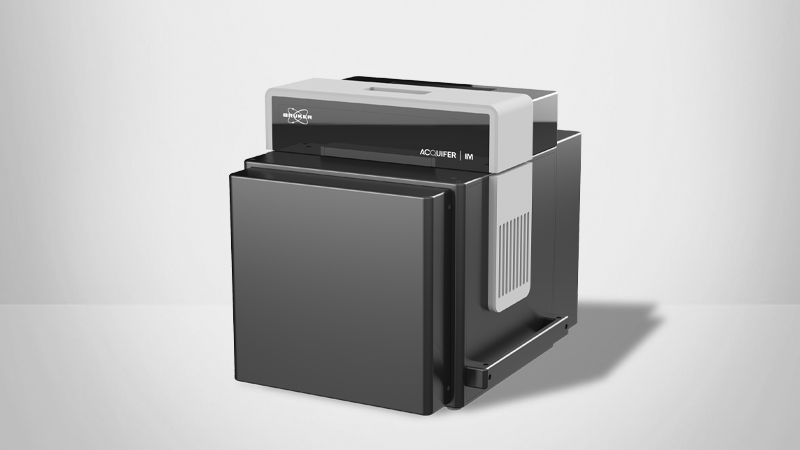
Tech Note: Remote Control and Unsupervised Feedback Microscopy with the Acquifer IM
Automated decision-making for complex high-content screening experiments
In this technical note, readers will learn about the features of the Acquifer IM that make it a preferred solution for unsupervised feedback microscopy including high-content screenings. The Acquifer IM includes a smart imaging interface and a built-in script editor for microscopy without the need for complex hardware communication protocols or third-party control software. This technical note will explain how Bruker's Acquifer supports automated decision-making for complex high-content screening experiments.
This technical note discusses:
- Typical difficulties associated with high-content screenings
- The features of Bruker's Acquifer Imaging Machine
- The benefits of smart imaging
- Requirements for unsupervised feedback microscopy
KEYWORDS: Acquifer Imaging Machine (IM), High-Content Screening, Unsupervised Feedback Microscopy, Remote Control, Smart Imaging, Imaging Workflow

Introduction
High-content screening is routinely employed to automatically acquire multi-dimensional image datasets at fixed positions within wells of microtiter plates. While this is sufficient for many applications, it imposes rather inflexible screening workflows as imaging positions are pre-defined by users, often regardless of specimen location or sample characteristics. This can lead to acquisition of unnecessary datasets, omission of features of interest, and could hinder more complex assays that would depend on real-time analysis of image data. Bruker's Acquifer Imaging Machine (IM) overcomes these challenges and enables unsupervised feedback microscopy and automated decision-making for complex high-content screening experiments.
Smart Imaging
Smart imaging describes methods for the semi-automated or automated recognition and analysis of structures of interest within samples, including the subsequent data acquisition from these regions. The analysis and decision-making are usually performed by external software packages that interface with the microscope’s software and hardware to remote control devices and modify imaging workflows depending on sample characteristics.
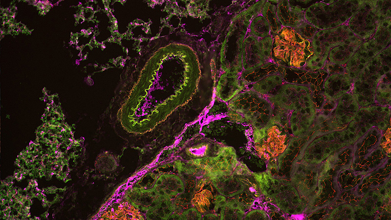
Smart imaging can massively improve higher magnification assays, as the limited field of view of higher magnification objectives often leads to omission of structures of interest at pre-defined imaging positions. Regions of interest (e.g. certain phenotypes) can be detected by external image processing routines and selectively imaged. This also enables tissue-specific imaging in large specimens (e.g. zebrafish), as the tissues or organ of interest can be automatically identified and imaged at high resolution. By restricting acquisition to interesting structures, smart imaging techniques save time, data volumes, and resources in large screening projects. Moreover, it enables complex assays like organ-specific screening in large specimen, which is not feasible by conventional high-content screening methods.

Acquifer IM's Smart Imaging Interface
Acquifer IM offers an easy-to-use smart imaging interface for unsupervised feedback microscopy. Via the Acquifer script language, virtually any external software (e.g. Fiji/ImageJ) that can produce text-based output can be readily interfaced with the screening microscope. This script-based communication between external software and imaging device enables unsupervised feedback microscopy without the necessity of complex hardware communication protocols or dedicated third-party control software.
Acquifer IM also includes an open software interface for developers that provides a built-in script editor for customization of imaging workflows, an easy-to-use file system-based interface to the Acquifer IM, and a TCP/IP interface for more advanced application developments. It also provides intuitive C#-based script language for command and control, the use of scripts for remote control, control imaging workflows, and hierarchical data structures.

Perform Unsupervised Feedback Microscopy with Ease
Acquifer IM is a system ideally equipped for smart imaging and enabling complex assays. It is adept at the semi-automated recognition and analysis of structures of interest within samples as well as the subsequent data acquisition from these regions. These capabilities have a significant impact on unsupervised feedback microscopy experiments and high-content screenings.
©2024 Bruker Corporation. Acquifer IM is a trademark of Bruker. All other trademarks are the property of their respective companies. All rights reserved. TN2605, Rev. A0.