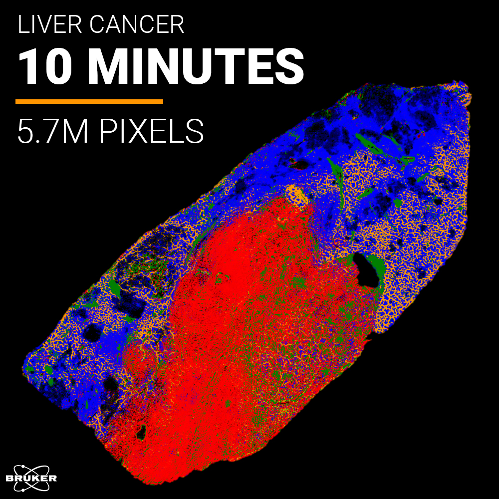
Tissue Analysis by Whole Slide IR Imaging
Automated Whole Slide IR Imaging - A World's First.
No staining, no labels, no waiting. TissuePlus™ is an end-to-end software workflow built for the LUMOS II ILIM that automates the entire workflow, from sample detection to data analysis, so you spend less time at the instrument and more time on your science.
Different tissues. Same easy workflow.
By choosing TissuePlusTM you get a complete, mature solution, that perform's tissue analysis at unprecedented speed and ease of use.
- Fully automated acquisition. Whole slide imaging runs hands-free from sample detection to data output, freeing researchers for other tasks.
- Rapid whole slide chemical analysis. Onboard plugins convert raw spectra into RGB integration overlays and segmentation maps the moment acquisition is complete, no manual post-processing required.
- Label-free and non-invasive. Tissue morphology and chemical composition is preserved, enabling reliable follow-up with histology or other imaging modalities.
- Reproducible results across operators. Consistent, high-quality data within and between large-scale studies — no IR expertise required.
- Open data format and Python API. Raw data is automatically stored in the open-source .zarr format, with a well-documented API for integrating custom algorithms as plugins.
How to Work With TissuePlusTM
Performing IR Laser Imaging of tissues has never been easier. See how TissuePlus™ automates the full workflow, from data acquisition to spectral analysis, delivering whole-slide results in minutes.
Routine Ready Workflow.
Define your measurement workflow once and reuse it across any number of samples. TissuePlus™ tracks every sample by status, keeps your parameters consistent across operators.

From Slide to Insight.
Press start and walk away. TissuePlus™ automatically detects tissue boundaries and starts data acquisition. The results are millions of full fingerprint IR spectra acquired in minutes. Once done, integrated analysis plugins process raw data into chemical images and clustering maps immediately. No manual steps between loading a slide and having results.

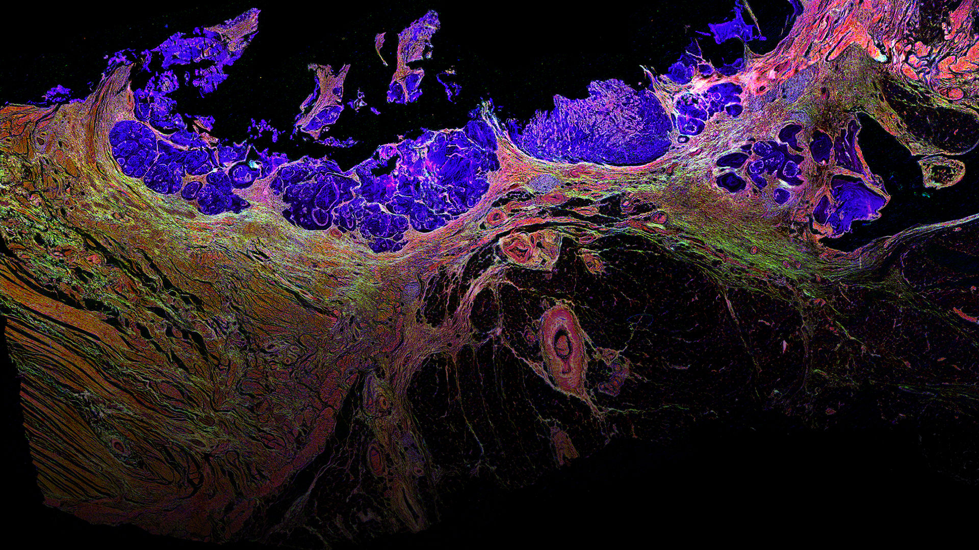
Get Morphological and Molecular Information in One Image.
Millions of IR spectra are automatically translated into reproducible, color-coded chemical images that render biochemical heterogeneity visible across entire sections. The same image simultaneously reveals tissue morphology, enabling confident identification of distinct phenotypes and pathological features.

Your data, your pipeline.
By storing raw data in the open-source .zarr format, TissuePlus™ ensures transparency, scalability, and interoperability. Its well-documented API supports the integration of user-defined algorithms and analysis pipelines. Teaching images enable straightforward alignment with complementary imaging modalities, like MALDI Imaging.
Shaped by our users, driven by science.
The unmet need for a streamlined whole slide IR imaging workflow came directly from our users. They clearly told us, they want to spend less time at the instrument, ensure robust operation, and achieve reproducible results across different operators. TissuePlus™ is our answer to that. Every design decision, from automated sample detection to onboard chemical analysis, reflects what users needed the most.

Bruker's ILIM’s speed of analysis is what we have been waiting for to use IR imaging to guide our
multimodal tissue analysis workflows. This opens up new avenues in many research projects.
Carsten Hopf, Ph.D., Director, Center for Mass Spectrometry and Optical Spectroscopy, Chair, Institute of
Instrumental Analytics and Bioanalytics, Professor of Bioanalytics and Drug Discovery, Mannheim University

We have been working in the field of tissue imaging and spectral pathology for over 20 years. One of the key barriers to clinical adoption is the long measurement times for full hyperspectral imaging of large areas of tissue. A sample set consisting of nearly 1500 prostate tissue cores, that took 3 months to measure on our old instrument, was measured in just two days on the Bruker ILIM system. This is a game changer!
Peter Gardner, Ph.D., Professor of Analytical and Biomedical Spectroscopy, University of Manchester