Application Note: BioAFM in the Cosmetic and Healthcare Industries
BioAFM in the Cosmetic and Healthcare Industries
Behind every new cream, serum, or therapeutic agent lies rigorous research and a complex development process aimed at enhancing efficacy, minimizing side effects, and ensuring regulatory compliance. Atomic force microscopy (AFM) delivers key insights in this process and is driving next-generation solutions in the cosmetic and healthcare industries.
This application note examines the capabilities of AFM and the benefits of its use in the healthcare and cosmetic sectors, and are illustrated with examples from a variety of applications. AFM enables an exact characterization of the structural, surface, and nanomechanical properties of additives, materials, and active ingredients, as well as a fast and easy assessment of product performance.
Readers can expect to learn about using BioAFM in the following examples:
- Contact lenses
- Dermatology and skincare
- Hair care
- Oral Care and dental protection
KEYWORDS: BioAFM, Atomic Force Microscopy, Cell Biology, Molecular Biology, Nanomechanical Mapping, AFM in Cosmetics, AFM in Healthcare

Humans have a long history of mixing ointments, balms, and salves for healing, protection, and cosmetic purposes. Cosmetic products, in particular, have long been an integral element of human culture, symbolizing care, self-confidence, and self-expression.1 Today, they combine dermatological science with aesthetic design, blending nourishing ingredients and advanced technologies for improved well-being and appearance. Behind every cream and serum, therapeutic agent, or medical device, such as contact lenses, lies rigorous research aimed at enhancing tolerance and performance, while minimizing adverse side effects. Atomic force microscopy (AFM) not only drives innovation and product development in the cosmetic and healthcare industries, it also serves as a powerful tool for quality control in routine production processes, ensuring consistent quality and reproducibility – essential factors for strengthening customer confidence and brand loyalty. This application note discusses how atomic force microscopes for biology (BioAFMs) can be used for a wide variety of applications in cosmetic and healthcare fields, and covers several examples in detail.
Introduction
AFM is a high-resolution microscopy technique that delivers three-dimensional images of the surface topography and a detailed mechanical characterization of samples, covering sub-nanometer to micrometer scales. AFM also enables the quantification of friction forces, adhesion, and viscoelastic properties, the identification of specific binding sites, and the investigation of dynamic processes. The ability of AFM to operate under near-physiological conditions, for example, at body temperature and under controlled humidity, makes it an ideal tool for investigating the effect of an active ingredient on living cells and tissues following application.2,3 In addition, the integration of artificial intelligence (AI) and deep learning algorithms is driving higher levels of automation, faster more precise measurements, and more comprehensive analyses.4
For companies that develop and manufacture healthcare products, medical devices, and cosmetics, these capabilities contribute towards the design of safer, more effective products and validate efficacy claims with quantitative data.5 By integrating AFM into development workflows, industries can better meet the growing regulatory and consumer demands for evidence-based quality – ensuring that products not only claim to be effective but are scientifically proven to be so.
LEARN MORE:
Applications: A Brief Overview
The following section presents examples of the use of AFM in the analysis of skin, hair, teeth, contact lenses, and oral care products. They illustrate the capabilities of AFM and the benefits of its use in the healthcare and cosmetics sector.
Contact Lenses
Contact lenses used for vision correction must meet multiple requirements simultaneously: optical performance, biocompatibility, and long-term comfort. These properties depend on a delicate interplay between material composition, surface topography, and mechanical properties. The nanoscale features of contact lenses strongly influence the stability of the tear film, wettability, and protein adsorption – critical factors for reducing irritation and the risk of infection.
As no single material can fully meet all of these requirements, modern contact lenses often combine optimized bulk materials with specialized surface coatings. AFM makes a powerful contribution towards the development of innovative lens materials in industrial research and quality assurance during the manufacturing process.6
High-resolution AFM imaging reveals surface roughness and heterogeneities in polymer structures, while nanomechanical mapping quantifies elasticity across the lens.7 In silicone hydrogel lenses, it is critical to balance oxygen permeability with surface smoothness.
Figure 1 summarizes representative AFM measurements on daily single-wear contact lenses, delivering:
- Large-area mapping of the lens curvature
- Stiffness measurements
- Nanoscale surface topography, and
- Viscoelastic properties
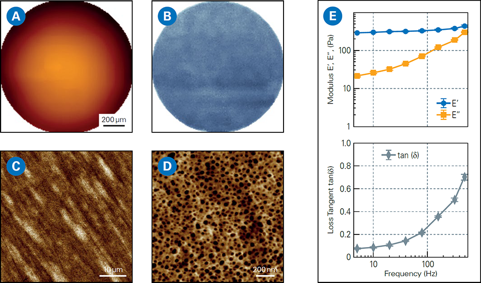
Advanced AFM modes, such as SmartMapping, perform large-area curvature mapping with nanoscale precision, enabling full-lens characterization at the millimeter scale.8
AFM measurements reveal key relationships between structure, composition, and performance, enabling a precise optimization of lens materials and design as well as quantifiable improvements in comfort, safety, and regulatory compliance.9,10
Research prototypes of next-generation lenses are exploring the integration of sensors for monitoring glucose or intra-ocular pressure, as well as augmented reality functionalities. AFM can play a vital role in this development, contributing towards precise material engineering and an exact characterization of material properties without compromising lens performance or wearer comfort.

Dermatology and Skincare
Skin health is an important indicator of overall wellbeing. Early detection of structural or compositional changes in the skin can significantly improve diagnostic and therapeutic outcomes. AFM enables the nanoscale characterization of skin by combining highresolution topographic imaging with nanomechanical mapping, in the so-called quantitative nanohistology.11
This approach provides objective biomarkers that go beyond the capabilities of conventional histology that typically relies on staining and light microscopy to assess tissue morphology and cellular architecture. AFM can detect subtle differences in tissue stiffness, elasticity, and surface morphology that often occur at the cellular or extracellular matrix level, before symptoms become visible at the macroscopic level.12 These nanoscale measurements are particularly valuable for distinguishing between healthy and diseased skin, in particular, in conditions like eczema, psoriasis, skin cancer, hypermobile Ehlers-Danlos syndrome (hEDS), or scleroderma.13,14
In a diagnostic study, AFM was used to examine dermal tissue sections from patients with hEDS and scleroderma. Two sections from the same sample were isolated, one was stained with Picosirius Red to visualize structural variations in the dermal collagen using polarized light microscopy, and the other was left unmodified and was analyzed using AFM. This approach allowed a direct correlation between classical histology and AFM-based nanomechanical analysis.
The AFM nano-topography analysis revealed distinct structural differences (see Figure 2A). Healthy skin displayed organized collagen fibrils with clear D-banding, while diseased regions showed chaotic arrangements of bent, randomly oriented, and twisted fibrils. In addition, AFM-derived biomechanical measurements exhibited significantly lower modulus values in hEDS-scleroderma tissue, indicating reduced collagen stiffness and greater structural disorder. In contrast, healthy skin showed a consistent, unimodal stiffness distribution with minor evidence of fibril irregularities (Figure 2B).

In addition to pathology, AFM can also assess hydration-induced swelling and lipid organization, delivering insights into the skin barrier function and irritation susceptibility.15,16 AFM’s ability to operate under physiologically relevant conditions makes it suitable for ex vivo biopsies and other promising future applications. By providing structural and biomechanical insights, AFM can enhance diagnoses, monitor disease progression, and support innovative developments in the field of skincare and cosmetics. AFM delivers quantitative metrics that enable the precise assessment of collagen structure, hydration, and the effects of treatment, supporting personalized skincare, targeted therapies, and scientifically backed product claims.
Human Hair
Human hair is a complex, keratin-based, anisotropic biomaterial that displays significant individual variability in thickness, curl, stiffness, and moisture. Its structural integrity determines its visual appearance, mechanical resilience, and tactile feel. Conventional evaluation methods – expert panel tests and macroscopic experiments – capture user perception but lack the ability to quantify nanoscale effects, such as adhesion, friction, and cuticle wear, factors that strongly influence the feel and durability of hair.17
The tribological properties of hair – the interplay between adhesion, friction and wear – are to a large extent dominated by the hair cuticle, the outmost protective layer of hair that is comprised of overlapping keratin scales oriented along the direction of hair growth. AFM is particularly suitable for the quantitative investigation of these properties and the evaluation of the performance of hair-care products.18-20
Figure 3 presents tribological AFM measurements performed with a 1 μm spherical probe along a hair fiber before (untreated) and after applying a conditioning hair care product. Although the corresponding topography images do not reveal clear visual differences between the surfaces of the hairs other than minor contaminants, the friction forces decrease significantly after treatment with a conditioner.
Transferring the friction force to the coefficient of friction according to Amonton’s law gives a scalable, quantitative parameter that can be linked to the performance of cosmetic products.18,19 Furthermore, the sliding speed can be varied to study dynamic tribology.20
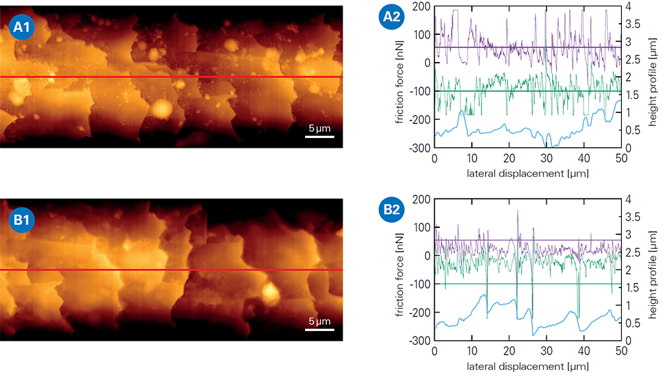
The automated workflows of AFM allow high-throughput analysis across numerous hair fibres and treatments. The design of the experiment can be adapted to mimic realistic conditions by mounting a segment of a hair fiber onto the cantilever, enabling the study of hair-to-hair contact.21 To simulate fingertip touch, specialized collagen-coated colloidal probes can be used to perform tribological measurements under conditions that reflect everyday handling.
BioAFM systems and their complementary environmental add-ons have been designed to operate in liquids and under controlled temperature and humidity conditions. This allows the analysis of shampoos, conditioners, and other cosmetic products under shower-like and everyday conditions, providing insights into their performance and effects on hair vitality, nutrition, and cuticle protection.22,23
Oral Care and Dental Protection
Oral health and, in particular, teeth exemplify the strong connection between aesthetic appearance and overall healthcare. White teeth are widely perceived to be a sign of health and social status, which explains the popularity of whitening products as a cosmetic treatment. Maintaining dental health, i.e., preventing dental decay (caries) and managing dentin hypersensitivity are key healthcare priorities.
Modern oral hygiene products, like toothpaste, are multifunctional, addressing both protective and whitening needs. Effective whitening requires bleaching agents that can compromise the integrity of the teeth and increase hypersensitivity by exposing dentin tubules.24,25 Oral care products must, therefore, balance cleaning and brightening with enamel protection.
AFM supports the development of such products by characterizing the surfaces of teeth at both the micro- and nanoscale level and under conditions that closely mimic reality. AFM quantifies surface roughness, mineral crystallite orientation, and micro-defects, properties that are critical for understanding how abrasives, bleaching agents, and biofilms affect the structure of teeth.26-28 This is essential for evaluating structural integrity and predicting the long-term performance of oral care treatments.
Figure 4 shows the effect of a bleaching agent on the surface structure of dentine, as observed by AFM. The noticeable changes to the surface structure after application demonstrate the aggressive properties of the bleaching agent.


Desensitizing toothpastes help reduce dentine hypersensitivity by, for example, incorporating ingredients that seal dentin tubule openings. In Figure 5, AFM was used to measure the effect of a desensitizing toothpaste. In addition, by examining multiple tubule openings, AFM can assess the effectiveness of the toothpaste by quantifying the proportion of sealed dentin tubules.
Nanomechanical mapping measures local stiffness and hardness, enabling direct assessment of enamel erosion or remineralization after treatment with fluoride or calcium-based formulations. In particular, contact resonance AFM can deliver high-resolution mechanical maps of very stiff surfaces, such as enamel and dentin, with an accuracy significantly beyond what conventional analysis methods can achieve.29
LEARN MORE:
Figure 6 presents a high‑resolution mechanical map of the dentin-enamel junction obtained using contact‑resonance AFM on the cross section of a human milk tooth. In addition to the topographic information, the difference in stiffness between enamel and dentin is clearly visible and corresponds to values reported in the literature.
These results highlight the ability of AFM to study chemical erosion and assess dental care treatments to provide a comprehensive evaluation of dental integrity. In addition, tribological analysis, performed under simulated brushing conditions, could optimize the development of dental hygiene products and formulations by, for example, determining the effects of the size of abrasive particles on performance to ensure effective cleaning and minimal enamel erosion. Similarly, advanced AFM modes can aid the design and evaluation of protective coatings that reduce bacterial adhesion and biofilm formation, critical factors for maintaining long-term oral health and preventing caries.
These approaches highlight the role AFM can play in guiding the development of nextgeneration oral care products that combine cosmetic benefits with oral healthcare.

Key Benefits of AFM in the Cosmetic and Healthcare Industries
Versatility
AFM delivers nanoscale insights into surface structure and mechanics across a diverse array of samples ranging from contact lenses to skin, hair, teeth, and care products, supporting innovation in both the cosmetic and healthcare industries.
Product Performance
High-resolution imaging and nanomechanical mapping enable the precise evaluation of material integrity, wear resistance, and the effects of treatments, ensuring optimized formulations and reliable quality control.
Realistic Testing
BioAFMs operate under physiologically relevant conditions, allowing the ex vivo analysis of samples, materials, and healthcare products before, during, and after application, enabling an accurate and comprehensive assessment of performance.
Advanced Tools
Techniques, such as contact resonance AFM and SmartMapping, deliver quantitative, multiparametric data that significantly surpass the results achieved by conventional analysis methods. The growing use of machine learning and AI-driven automation in AFM is rapidly accelerating workflows and improving reproducibility.
Future Potential
AFM is expected to drive next-generation solutions, from smart contact lenses to personalized skincare and advanced oral care coatings, while assisting manufacturers to meet regulatory and consumer demands for evidence-based performance.
Authors
Maximilian Seuss, Jörg Barner, Randhir Kumar, and Thomas Henze.
For more information, please contact us at support.bioafm@bruker.com.
References
- McMullen RL, Dell’Acqua G (2023). History of Natural Ingredients in Cosmetics. Cosmetics 10:71. https://doi.org/10.3390/cosmetics10030071
- Alsteens D (2024). Probing living cell dynamics and molecular interactions using atomic force microscopy. Biophys Rev 16:663–677. https://doi.org/10.1007/s12551-024-01258-7
- Magazzù A, Marcuello C (2023). Investigation of Soft Matter Nanomechanics by Atomic Force Microscopy and Optical Tweezers: A Comprehensive Review. Nanomaterials 13:963. https://doi.org/10.3390/nano13060963
- Di Guardo A, Trovato F, Cantisani C, Dattola A, Nisticò SP, Pellacani G, Paganelli A (2025). Artificial Intelligence in Cosmetic Formulation: Predictive Modeling for Safety, Tolerability, and Regulatory Perspectives. Cosmetics 12:157. https://doi.org/10.3390/cosmetics12040157
- Bhushan B, LaTorre C (2008). Structural, Nanomechanical, and Nanotribological Characterization of Human Hair Using Atomic Force Microscopy and Nanoindentation. In: Nanotribology and Nanomechanics. Springer Berlin Heidelberg, Berlin, Heidelberg, pp 1325–1485
- Last JA, Russell P, Nealey PF, Murphy CJ (2010). The Applications of Atomic Force Microscopy to Vision Science. Invest Ophthalmol Vis Sci 51:6083. https://doi.org/10.1167/iovs.10-5470
- Sharma V, Shi X, Yao G, Pharr GM, Wu JY (2022). Surface characterization of an ultrasoft contact lens material using an atomic force microscopy nanoindentation method. Sci Rep 12:20013. https://doi.org/10.1038/s41598-022-24701-9
- Bruker Nano Surfaces SmartMapping. In: AFM Modes - SmartMapping. https://www.bruker.com/en/products-and-solutions/microscopes/bioafm/bioafm-modes/smartmapping.html
- Mitrovic A, Bojovic B, Stamenkovic D, Popovic D (2018). Characterization of surface roughness of new nanophotonic soft contact lenses using lacunarity and AFM method. Hem Ind 72:157–166. https://doi.org/10.2298/HEMIND170924004M
- Kim SH, Lee J, Jang YJ, Lee K-B, Lee Y (2020). ToF-SIMS and AFM Characterization of Brown Cosmetic Contact Lenses: From Structural Analysis to the Identification of Pigments. Journal of Analytical Methods in Chemistry 2020:1–11. https://doi.org/10.1155/2020/6134627
- Huang S, Strange A, Maeva A, Siddiqui S, Bastien P, Aguayo S, Vaez M, Montagu-Pollock H, Ghibaudo M, Potter A, Pageon H, Bozec L (2023). Quantitative nanohistology of aging dermal collagen. Front Aging 4:1178566. https://doi.org/10.3389/fragi.2023.1178566
- He T, Fisher GJ, Kim AJ, Quan T (2023). Age-related changes in dermal collagen physical properties in human skin. PLoS ONE 18:e0292791. https://doi.org/10.1371/journal.pone.0292791
- Kotova SL, Timashev PS, Guller AE, Shekhter AB, Misurkin PI, Bagratashvili VN, Solovieva AB (2015). Collagen Structure Deterioration in the Skin of Patients with Pelvic Organ Prolapse Determined by Atomic Force Microscopy. Microsc Microanal 21:324–333. https://doi.org/10.1017/S1431927615000148
- Neshatian M, Mittal N, Huang S, Ali A, Khattignavong E, Bozec L (2024). Investigation of dermal collagen nanostructures in Ehlers-Danlos Syndrome (EDS) patients. PLoS ONE 19:e0307442. https://doi.org/10.1371/journal.pone.0307442
- Qin X, Dong M, Li Q (2024). Insight into the hydration friction of lipid bilayers. Nanoscale 16:2402–2408. https://doi.org/10.1039/D3NR05517E
- Marcott C, Lo M, Kjoller K, Domanov Y, Balooch G, Luengo GS (2013). Nanoscale infrared (IR) spectroscopy and imaging of structural lipids in human stratum corneum using an atomic force microscope to directly detect absorbed light from a tunable IR laser source. Experimental Dermatology 22:419–421. https://doi.org/10.1111/exd.12144
- Lodge RA, Bhushan B (2006). Surface characterization of human hair using tapping mode atomic force microscopy and measurement of conditioner thickness distribution. Journal of Vacuum Science & Technology A: Vacuum, Surfaces, and Films 24:1258–1269. https://doi.org/10.1116/1.2180275
- Smith JR, Tsibouklis J, Nevell TG, Breakspear S (2013). AFM friction and adhesion mapping of the substructures of human hair cuticles. Applied Surface Science 285:638–644. https://doi.org/10.1016/j.apsusc.2013.08.104
- Bowen J, Johnson SA, Avery AR, Adams MJ (2016). Friction and wear of human hair fibres. Surf Topogr: Metrol Prop 4:024008. https://doi.org/10.1088/2051-672X/4/2/024008
- Weiand E, Ewen JP, Roiter Y, Koenig PH, Page SH, Rodriguez-Ropero F, Angioletti-Uberti S, Dini D (2023). Nanoscale friction of biomimetic hair surfaces. Nanoscale 15:7086–7104. https://doi.org/10.1039/D2NR05545G
- Luengo GS, Mizuno H, Rutland MW (2017). Hair-Hair Contact Dynamics and Interactions Studied with Atomic Force Microscopy. In: Humbert P, Fanian F, Maibach HI, Agache P (eds) Agache’s Measuring the Skin. Springer International Publishing, Cham, pp 835–845
- Fellows AP, Casford MTL, Davies PB (2020). Nanoscale Molecular Characterization of Hair Cuticle Cells Using Integrated Atomic Force Microscopy–Infrared Laser Spectroscopy. Appl Spectrosc 74:1540–1550. https://doi.org/10.1177/0003702820933942
- Fellows AP, Casford MTL, Davies PB (2022). Chemically characterizing the cortical cell nano-structure of human hair using atomic force microscopy integrated with infrared spectroscopy (AFM-IR). Intern J of Cosmetic Sci 44:42–55. https://doi.org/10.1111/ics.12753
- Dahl JE, Pallesen U (2003). Tooth bleaching - a critical review of the biological aspects. Critical Reviews in Oral Biology & Medicine 14:292–304. https://doi.org/10.1177/154411130301400406
- De Freitas MR, De Carvalho MM, Liporoni PCS, Fort ACB, Moura RDME, Zanatta RF (2021). Effectiveness and Adverse Effects of Over-the-Counter Whitening Products on Dental Tissues. Front Dent Med 2:687507. https://doi.org/10.3389/fdmed.2021.687507
- Pattem J, Field J, Waterhouse PJ, German MJ (2022). The dynamic interplay of dietary acid pH and concentration during early-stage human enamel and dentine erosion. Front Dent Med 3:1040565. https://doi.org/10.3389/fdmed.2022.1040565
- Murchie BD, German MJ, Waterhouse PJ, Mullan F (2025). The early wear susceptibility of dentine following exposure to citric acid: In vitro study. Archives of Oral Biology 178:106363. https://doi.org/10.1016/j.archoralbio.2025.106363
- Zhai Y, Shi Z, Premaraj T, Premaraj S, Karpova T, Dong P, Gu L (2025). Effects of Acid Etching on the Microstructure and Stiffness of Human Teeth. Journal of Engineering and Science in Medical Diagnostics and Therapy 8:031005. https://doi.org/10.1115/1.4066624
- Bruker Nano Surfaces Contact Resonance Module. In: BioAFM Accessories and Add-Ons - Contact Resonance Module. https://www.bruker.com/en/products-and-solutions/microscopes/bioafm/bioafm-accessories/contact-resonance-module.html