Application Note: Correlative AFM and Super-Resolution STED Analysis of DNA Nanostructures
Recent developments in high-speed AFM imaging (>10 fps) and the development of advanced imaging and quantitative property mapping modes have greatly expanded our ability to characterize kinetic and dynamic processes as well as very soft, very small, and fragile structures like single molecules in liquid.
The NanoWizard® ULTRA Speed 2* with the Vortis™ controller can be coupled to a super-resolution STED instrument. This setup leverages the advantages of both techniques to enable true correlative microscopy and non-invasive examination of specific molecules or features carrying immunochemical information.
In this application note, we exhibit the capability of the NanoWizard ULTRA Speed instrument to non-invasively scan DNA nanostructures at 400 lines/s in closed-loop mode. We demonstrate that the instrument can:
- Be successfully coupled to a 2D compact line super-resolution STED instrument;
- Non-invasively carry out correlative microscopy with lateral resolution beyond 70 nm; and
- Image dynamic processes with a temporal resolution of over 1 fps.
*The latest generation system in this product line is the NanoWizard ULTRA Speed 3
Readers can expect to learn about:
- How and why to use the NanoWizard ULTRA Speed instrument to study DNA-based nanostructures with sub-molecular resolution in vitro.
KEYWORDS: Fast Scanning AFM; Correlative Microscopy; DNA Nanorulers; DNA Origami; High-Resolution Imaging; STED; Super-Resolution Imaging
Introduction
In the past decades, atomic force microscopy (AFM) has become standard in the high-resolution structural analysis of samples ranging from single molecules to complex macromolecular systems 1, 2. Unlike other high-resolution imaging techniques, it does not require any specific sample modification (except for surface deposition), the risk, therefore, of introducing artefacts through sample preparation is reduced. In recent years, there has been an increased demand for and application of novel developments featuring high-speed AFM imaging (>10 frames/sec), quantitative nanomechanical characterization of samples, and advanced feedback imaging modes which tweak the maximum attainable resolution.
Furthermore, the examination of, e.g. specific molecules or features carrying immunochemical information has been made possible by combining AFM with recent developments in optical/fluorescence microscopy, and in doing so, complementing the advantages of both techniques and enabling true correlative microscopy 3.
Correlative and high-speed AFM
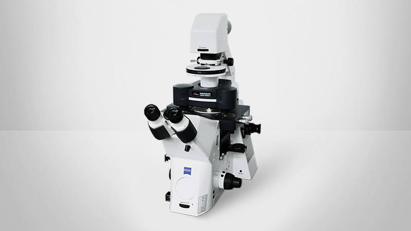
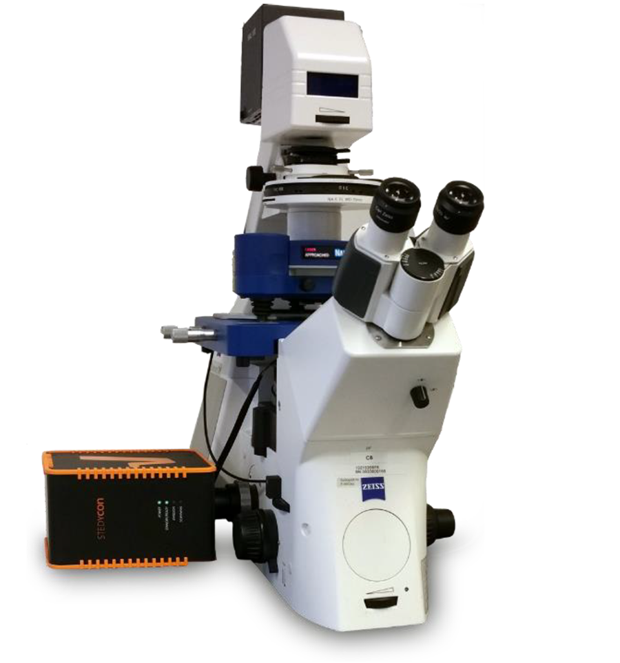
The measurements presented here were carried out using the recently launched NanoWizard® ULTRA Speed 2 with the 2nd generation Vortis™ (Bruker Nano), a benchmark in correlative, high-speed BioAFM combined with advanced optical microscopy. The system features easy-to-use modes such as QI™, which allows reliable and reproducible imaging of very soft and delicate samples in liquid, as well as the quantification of mechanical and adhesive sample properties with nanometer resolution. More advanced modes such as the HyperDrive™ allow highest resolution imaging of the topography of very small and fragile structures like single molecules in liquid. To conclude, we have demonstrated the capability of the instrument to non-invasively scan DNA nanostructures at 400 lines/s in closed-loop mode.
A schematic representation of a typical instrumental setup for the advanced correlative measurements used here is shown below (Figure 1).
STED - a super-resolution microscopy technique
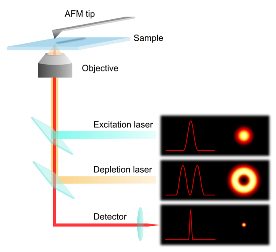
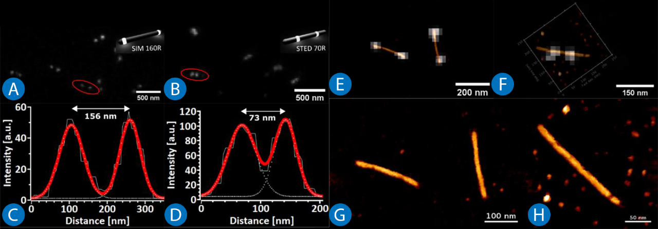
In recent years a variety of super-resolution microscopy techniques have been developed, all proposing different concepts for overcoming the Abbe diffraction limit, which defines the minimum resolvable distance between two point-like objects 4. The resolution of an optical microscope ends at approx. 200 nm laterally and 600 nm axially. In 1994, Hell and Wichmann developed a unique strategy for breaking the diffraction limit, stimulated emission depletion (STED), which was awarded the Nobel Prize in Chemistry in 2014 5. STED microscopy uses two laser pulses, the first one excites and the second one de-excites the fluorophores, fluorescent chemical compounds
that can re-emit light upon light excitation. The excitation laser focus scans the sample, whereas the depletion donut-shaped STED beam switches off the fluorophores at the rim of the focal point, thus confining the spot of effective fluorescence (Figure 2). Increasing the intensity of the STED beam reduces the full width at half maximum (FWHM) of the effective focal spot dSTED as shown below:

with λ being the wavelength, n - refractive index of the medium, a - the half-aperture angle of the objective, I depletion STED beam intensity, and IS - characteristic saturation intensity for a particular fluorophore at which the emitted fluorescence is reduced by a factor of 1/e.
DNA as a nanoscale building block
Deoxyribonucleic acid (DNA) is a polymer that chemists dream of. Its inherent structure is based on four organic bases that comprises two alpha-helical strands held together by hydrogen bonds 6. Following a certain complementarity code, the sequences in the strands not only bind to one another, but also create numerous possibilities for (non)-directed self-assembly into intricate shapes and structures on the nanoscale 7. The sequence of a DNA strand defines the way it assembles, which allows the prediction/modelling of the final structure a DNA molecule will adopt in vitro. By using specific sequences, one or several DNA strands can be forced to assemble into complex architectures. Seeman laid the foundation for exploiting DNA for nanotechnology and described several strategies for the formation of complex DNA structures 8. The ongoing development of novel design strategies increases the diversity of structures and their applications in many fields. For instance, Ackermann et al. presented rotaxanes made from DNA, opening new possibilities for research into molecular machines and synthetic biology 9, 10. Benson et al. demonstrated an automated technique for converting digitally generated flat sheet meshes into scaffolded DNA structures 11.
DNA-based nanorulers
Another application is the use of DNA origami structures as calibration standards for 2D and 3D super-resolution optical microscopes 12-14. Commercially available DNA-based rulers, used for the quantification and calibration of super-resolution systems and with a precision of just a few nanometers, are available in different forms with varying fluorophore spacings, fluorescence tags, as well as nanostructure shape and size 15. To demonstrate the compatibility of such structures for correlative AFM measurements, we used commercially available STED nanorulers (GATTAquant GmbH), which are rod-shaped and modified with dye molecules placed at defined distances along the rod (Figure 3 a, b).
DNA-based origami
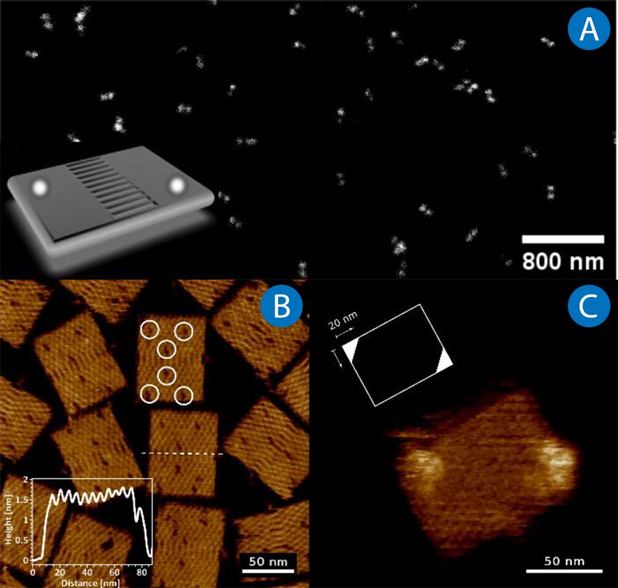
In certain scenarios, it is advantageous to use both an optical and an AFM calibration standard. In this work, we used a new rectangular origami (NRO) design with the dimensions 100 x 70 nm 14. Such structures are commercially available as GATTA-AFM, in which case they selectively carry two diagonally spaced Atto647N fluorophore populations (Figure 4a - inset). The STED signal from such structures is shown in Figure 4a. The molecular substructure of such conformations is easily recognizable in AFM images due to their planar design and two characteristic hallmarks of their pattern (Figure 4c). The first is the central ladder seam bridging the crossed halves of the design, which has a pitch of 6 nm, as stated in 15. Secondly, there are six visible gaps in the NRO lattice (labelled here with circles), which are introduced via shortened staple strands, as designated binding sites for other molecules 14. The NRO design carrying fluorophores was analysed with phase-modulation AFM (HyperDriveTM) and is shown here in Figure 4c. According to the manufacturer, each binding locus (20 x 20 nm) on the lattice is hybridised by 12-15 ssDNA. These locations are clearly visible at each end, however, due to their length (25 bp), they exhibit a certain hydrodynamic radius fluctuation which prevents a clear resolution of the NRO lattice.
High-speed imaging of DNA origami
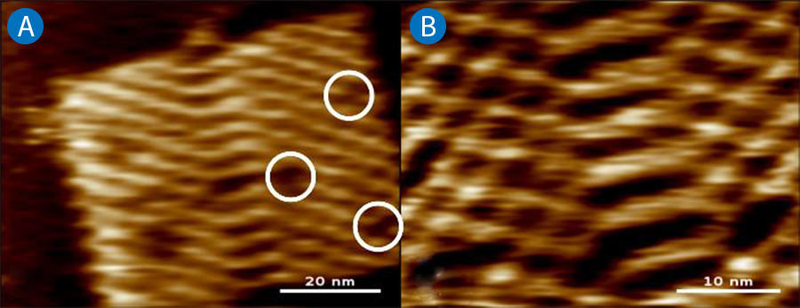
High-speed applications are an ongoing development in the repertoire of AFM and have been found to be particularly interesting in the characterization of kinetic and dynamic processes. In the case of DNA origami, such events could be the (non-)assisted self-assembly of surface structures and lattices, or simply the binding of specific single molecules and cargo. Here, we studied the NRO lattices (without fluorophores) with high-speed AFM (Figure 5).
The resulting AFM images show that it is possible to non-invasively study the molecular structures of the NRO at a line rate of 400 lines/s and with an image resolution of 256 x 256 pixels. The temporal resolution of 1.6 frames/s (fps) can be further increased to over 10 fps by applying the bi-directional scanning implemented in the NanoWizard ULTRA Speed 2, and/or by scaling down the line resolution. This would in turn make it possible to resolve dynamic events taking place on the millisecond scale.
Conclusion
We have successfully used the latest generation NanoWizard ULTRA Speed 2 AFM to study DNA-based nanostructures with sub-molecular resolution in vitro. Furthermore, it was demonstrated that the instrument can be successfully coupled to a 2D compact line super-resolution STED instrument and can, non-invasively, carry out correlative microscopy with a lateral resolution better than 70 nm. Furthermore, the ability of this setup to study dynamic processes with a temporal resolution of over 1 fps was also demonstrated.
Acknowledgements
All nanoruler & NRO samples were kindly provided by GATTAquant GmbH, Hiltpoltstein, Germany. The Stedycon compact line STED was kindly provided by Abberior Instruments GmbH, Göttingen, Germany.
Authors
Tanja Neumann, Jörg Barner, Dimitar Stamov (JPK BioAFM Business, Bruker Nano GmbH)
References
- Stamov DR, Kaemmer SB, Hermsdörfer A, Barner J, Jähnke T, Haschke H (2015) BioScience AFM Capturing Dynamics from Single Molecules to Living Cells. Microsc Today 23:18 25 . doi: 10.1017/S1551929515001005
- Winkel AK, Barner J, Henze T, Neumann T, Körnig A, Kumpfe F, Haschke H (2016) The Wide-Open Door: Atomic Force Microscopy 30 Years On. Microsc Today 24:12 17 . doi: 10.1017/S1551929516000900
- Monserrate A, Casado S, Flors C (2014) Correlative Atomic Force Microscopy and Localization-Based Super-Resolution Microscopy: Revealing Labelling and Image Reconstruction Artefacts. ChemPhysChem 15:647 650 . doi: 10.1002/cphc.201300853
- Endesfelder U (2018) Super-Resolution Microscopy. A Practical Guide. By Udo J. Birk. Angew Chem Int Ed 57:7939 7939 . doi: 10.1002/anie.201804434
- Hell SW, Wichmann J (1994) Breaking the diffraction resolution limit by stimulated emission: stimulated-emission-depletion fluorescence microscopy. Opt Lett 19:780 782 . doi: 10.1364/OL.19.000780
- Watson JD, Crick FHC (1953) Molecular Structure of Nucleic Acids: A Structure for Deoxyribose Nucleic Acid. Nature 171:737 738 . doi: 10.1038/171737a0
- Sanderson K (2010) Bioengineering: What to make with DNA origami. Nature 464:158 159 . doi: 10.1038/464158a
- Seeman NC (1998) DNA NANOTECHNOLOGY: Novel DNA Constructions. Annu Rev Biophys Biomol Struct 27:225 248 . doi: 10.1146/annurev.biophys.27.1.225
- Ackermann D, Schmidt TL, Hannam JS, Purohit CS, Heckel A, Famulok M (2010) A double-stranded DNA rotaxane. Nat Nanotechnol 5:436 442 . doi: 10.1038/nnano.2010.65
- Ackermann D, Jester S-S, Famulok M (2012) Design Strategy for DNA Rotaxanes with a Mechanically Reinforced PX100 Axle. Angew Chem Int Ed 51:6771 6775 . doi: 10.1002/anie.201202816
- Benson E, Mohammed A, Bosco A, Teixeira AI, Orponen P, Högberg B (2016) Computer-Aided Production of Scaffolded DNA Nanostructures from Flat Sheet Meshes. Angew Chem Int Ed 55:8869 8872 . doi: 10.1002/anie.201602446
- Steinhauer C, Jungmann R, Sobey T, Simmel F, Tinnefeld P (2009) DNA Origami as a Nanoscopic Ruler for Super-Resolution Microscopy. Angew Chem Int Ed 48:8870 8873 . doi: 10.1002/anie.200903308
- Schmied JJ, Forthmann C, Pibiri E, Lalkens B, Nickels P, Liedl T, Tinnefeld P (2013) DNA Origami Nanopillars as Standards for Three-Dimensional Superresolution Microscopy. Nano Lett 13:781 785 . doi: 10.1021/nl304492y
- Schmied JJ, Raab M, Forthmann C, Pibiri E, Wünsch B, Dammeyer T, Tinnefeld P (2014) DNA origami based standards for quantitative fluorescence microscopy. Nat Protoc 9:1367 1391 . doi: 10.1038/nprot.2014.079
- Gattaquant DNA Nanotechnologies. In: Gattaquant DNA Nanotechnologies. http://www.gattaquant.com/. Accessed 28 Jan 2019
SEE THE LATEST GENERATION NANOWIZARD ULTRA SPEED: