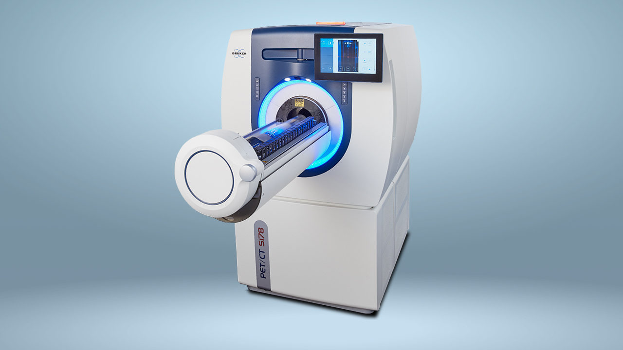
Multi-modal PET Technology Provides Novel Insights in Preclinical Oncology
Imaging Cancer: Multi-modal PET Technology Provides Novel Insights in Preclinical Oncology
Todd Sasser, Head of PCI Applications, Americas & Snr NMI Applications Scientist, Bruker BioSpin
Positron Emission Tomography (PET) is a widely-used imaging technique in nuclear medicine, for both clinical diagnostics and preclinical applications. PET provides three-dimensional (3D) functional imaging using radioactive tracers (radiotracers), showing the spatial distribution of biomolecular activity in the bodies of animal models and humans. Small animal studies are necessary to explore and validate imaging agents in the preclinical phase, before beginning clinical trials. In this context, PET imaging is increasingly being used by researchers in the drug development phase as it provides data that can be extrapolated from animal to human studies. Preclinical imaging facilitates the development and evaluation of novel therapeutic strategies, by providing important insights into disease mechanisms, from molecular to organ level. Small animal imaging deepens our understanding of disease development and the effect of potential treatments, and advances in PET technology are powering the translation of this res arch into the clinical setting.
The need to offer patients more personalized cancer treatment is driving advances in preclinical PET oncology research. The numerous different types of tumors – including those not yet well characterized – and their varying reactions to treatment, make the search for new effective cancer therapies incredibly challenging. Non-invasive in vivo imaging technologies such as PET enable researchers to better understand the course of tumor progression, by visualizing cancer-related processes in real-time. The combination of other imaging modalities, such as magnetic resonance imaging (MRI), computed tomography (CT), and single-photon emission computed tomography (SPECT), joins structural and functional imaging in one experiment. PET/CT, PET/MR and PET/SPECT/CT multi-modal systems are able to provide quantitative 3D tomographic images of radiotracers, bone, and soft tissue, furthering the growing knowledge of cancer biology and treatment.
Download the full application note: