Tech Note: Real-Time Visualization of Biomolecular Dynamics with the NanoRacer High-Speed AFM
Real-Time Visualization of Biomolecular Dynamics with the NanoRacer High-Speed AFM
Observing the structure, dynamics, and function of biological samples, and understanding the intrinsic relationship between them, has become a key focus of modern biophysics and molecular biology. In particular, there is a growing demand for tools that enable the investigation of the dynamics of individual biomolecules under conditions that closely mimic their native physiological environment.
This application note examines the capabilities of high-speed atomic force microscopy (HS-AFM), a powerful technique that enables the measurement of the kinetics of molecular processes and structural dynamics of biomolecules with high spatio-temporal resolution and minimal sample invasiveness.
Readers can expect to learn about:
- An outline of recent technical advancements
- Recommendations for achieving optimum performance
- A brief overview of novel applications using HS-AFM
KEYWORDS: High-speed Atomic Force Microscopy; HS-AFM; Biomolecules; DNA; Single-molecules; Kinetics; Dynamics; molecular mechanisms

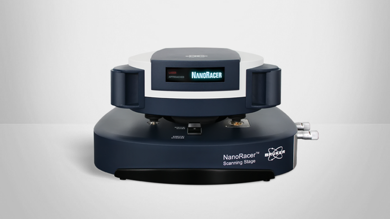
Observing the structure, dynamics, and function of biological samples, and understanding the intrinsic relationship between them, has become a key focus of modern biophysics and molecular biology. In particular, there is a growing demand for tools that enable the investigation of the dynamics of individual biomolecules under conditions that closely mimic their native physiological environment. High-speed atomic force microscopy (HS-AFM) is a powerful technique that enables the measurement of the structural dynamics of biomolecules with high spatio-temporal resolution and minimal sample invasiveness.1 Recent progress in the availability of probes suitable for high-speed applications and technical advances made to critical AFM components have significantly enhanced the ability to capture the dynamics of biological processes, which often occur in the millisecond range, in unprecedented detail.2 This technical note discusses how Bruker’s award-winning NanoRacer® High-Speed AFM3 enables the investigation of molecular dynamics at sub-20-ms scale for novel applications and gives a brief overview of a case study that demonstrates how highspeed atomic force microscopy can be used in the real-time visualization of DNA dehybridization and rehybridization.

NanoRacer High-Speed AFM Advancements
Several key technical innovations in the NanoRacer provide unique capabilities and outstanding performance. These include three-axis, closed-loop, sample scanning, a high-bandwidth amplifier, and sophisticated scanner and feedback control algorithms to enable:
- Video-rate scanning speeds of up to 50 frames per second (fps), and
- Optional photothermal probe excitation for highly stable cantilever excitation over extended periods.
NanoRacer’s user-friendly hardware and software are easy to operate, even for beginners. Features include:
- A sample scanner designed to measure samples in a liquid environment,
- An AFM head and scanner that can be easily removed from the stage for convenient sample processing, and
- A specialized, sealed liquid cell for experiments with very small volumes (down to 30 µl), precise control of environmental conditions and the concentration of, e.g., chemicals and proteins, or the exchange of buffers.
Achieving Optimum Performance
To get the best results when performing high-speed atomic force microscopy imaging, the biomolecules must be attached to a smooth substrate with minimal surface roughness. This ensures the accurate identification of the specific molecules being studied. In addition, the immobilization should be weak enough not to impair the dynamics of the molecules. Two critical factors are:
- Functionalization of the substrate, and
- Buffer conditions chosen with respect to the isoelectric point and surface charge of the molecules.
Typical surfaces suitable for high-speed atomic force microscopy imaging are:
- Mica for DNA, proteins, and lipids,
- Supported Lipid Bilayers (SLB) for individual membrane proteins, and
- DNA origami with specific binding sites for proteins.
With the correct selection of the appropriate substrate, imaging conditions, and functionalization, it is possible to stop diffusion and preserve the dynamic motions of individual molecules.
Wide Range of Applications
The NanoRacer High-Speed AFM opens the way for a host of new and innovative applications, such as the study of the structural dynamics of proteins, their intra- and inter-subunit interactions, and conformational changes. Some of the more notable studies include the real-time visualization of myosin V translocation along actin filaments,4 the rotational dynamics of annexin V trimers and their role in membrane healing,5 and the structural and conformational dynamics of intrinsically disordered proteins.6
An examplary application is the investigation of the dynamics of DNA rehybridization.
Previous studies have shown that pUC19 plasmids bind to polycationic interfaces, forming constructs with significant torsional energy,7,11 and are highly inclined to form dehybridization DNA bubbles (using single stranded regions of DNA) (Figure 2).
High-speed imaging of the samples revealed that these bubbles spontaneously open and close along the DNA double strand. Rezipping of the DNA double strand leads to healing of the DNA bubble, thus verifying the kinetics of the DNA dehybridization processes. By performing high-speed atomic force microscopy imaging at 40 frames per second, it was possible to visualize thermodynamic state fluctuations of single DNA strands and, ultimately, their multiple conformational states before rehydridization of the strands.
The continuously growing list of high-speed atomic force microscopy applications and their significance7-10 includes, but is not limited to the following experiments:

| Lipid Phase Transition | Capturing dynamic changes in lipid phases – provides insights into membrane fluidity and organization7 |
| Streptavidin-Biotin Binding Kinetics | Observation of binding interactions at the single-molecule level – crucial for understanding molecular recognition and binding dynamics7,8 |
| Thermodynamic DNA Rehybridization | Monitoring of de-/rehybridization of DNA strands – sheds light on the thermodynamics and kinetics of nucleic acid interactions7 |
| Collagen Type I Fibrillogenesis Kinetics | Tracking the assembly of collagen fibrils – crucial for understanding tissue formation and repair mechanisms7 |
| Ribonucleoprotein Assembly Dynamics | Visualization of the assembly of ribonucleoproteins – useful in the study of RNA-protein interactions and their role in cellular processes |
| αSyn Amyloid Formation Kinetics | Observing the formation of amyloid fibrils – essential for understanding the molecular origins of neurodegenerative diseases9 |
| Chromatin Recognition Module Bridging Activity | Investigation of how chromatin recognition modules interact and bridge DNA – provides insights into gene regulation and chromatin organization10 |
Outlook and Future Trends
The NanoRacer High-Speed AFM is a highly sensitive instrument that offers significant advantages for high-speed applications. It operates with exceptionally low noise levels and is an exceptionally stable system. Significant capabilities include minimal sample damage, clean cantilever oscillation via photothermal excitation, photothermal excitation, and advanced scanner control, making it ideal for long-term, highly accurate experiments. Future research in the field of single-molecule biology can leverage these features to explore biomolecular mechanisms, such as enzyme kinetics, protein dynamics, and molecular assembly processes.
Conclusions
Acknowledgements
The author would like to thank Dr. Andreas Kraus (formerly Bruker, currently TESVOLT AG) and Dr. Heiko Haschke (Bruker) for their assistance with the NanoRacer measurements. We would also like to thank Dr. Thomas Henze (Bruker) for the discussions and critical reviews during the preparation of the document.
Author
Dr. Prem Kumar Viji Babu (Bruker)
References
- Ando T, Uchihashi T, Scheuring S (2014), Filming Biomolecular Processes by High-Speed Atomic Force Microscopy, Chem Rev 114 (6), 3120–3188.
- Fukuda S, Ando T (2023), Technical advances in high-speed atomic force microscopy. Biophys Rev. https://doi.org/10.1007/s12551-023-01171-5
- Lyman C (2021), 2021 Microscopy Today Innovation Awards, Microsc Today 29 (5), 12–17.
- Kodera N, Yamamoto D, Ishikawa R, Ando T (2010), Video imaging of walking myosin V by high-speed atomic force microscopy, Nature 468 (7320), 72–76.
- Miyagi A, Chipot C, Rangl M, Scheuring S (2016), High-speed atomic force microscopy shows that annexin V stabilizes membranes on the second timescale, Nat Nanotechnol 11 (9), 783–790.
- Kodera N, Noshiro D, Dora SK, Mori T, Habchi J, Blocquel D, Gruet A, Dosnon M, Salladini E, Bignon C, Fujioka Y, Oda T, Noda NN, Sato M, Lotti M, Mizuguchi M, Longhi S, Ando T (2021), Structural and dynamics analysis of intrinsically disordered proteins by high-speed atomic force microscopy, Nat Nanotechnol 16 (2), 181–189.
- Stamov DR, Neumann T, Kraus A, Körnig A, Jankowski T, Knebel D, Jähnke T, Henze T, Haschke H (2022), Unraveling Molecular Dynamics with High-Speed Video-Rate Atomic Force Microscopy, Microsc Today 30 (3), 10–14.
- Domínguez CM, García-Chamé M, Müller U, Kraus A, Gordiyenko K, Itani A, Haschke H, Lanzerstorfer P, Rabe KS, Niemeyer CM (2022), Linker Engineering of Ligand-Decorated DNA Origami Nanostructures Affects Biological Activity, Small 18 (35), 2202704.
- Franco A, Gracia P, Colom A, Camino JD, Fernández-Higuero JÁ, Orozco N, Dulebo A, Saiz L, Cremades N, Vilar JMG, Prado A, Muga A (2021), All-or-none amyloid disassembly via chaperone-triggered fibril unzipping favors clearance of α-synuclein toxic species, Proc Natl Acad Sci 118 (36), e2105548118.
- Burdett H, Foglizzo M, Musgrove LJ, Kumar D, Clifford G, Campbell LJ, Heath GR, Zeqiraj E, Wilson MD (2023), BRCA1–BARD1 combines multiple chromatin recognition modules to bridge nascent nucleosomes, Nucleic Acids Res 51 (20), 11080–11103.
- Bruker Nano Surfaces White Paper (2019), High-speed imaging of DNA submolecular structure and dynamics. Microsc. Anal. https://analyticalscience.wiley.com/content/ news-do/high-speed-imaging-dna-submolecular-structure-and-dynamics
©2025 Bruker Corporation/Bruker Nano GmbH. NanoRacer is trademark of Bruker Nano GmbH. All other trademarks are the property of their respective companies. All rights reserved. TN302 Rev. A0.