
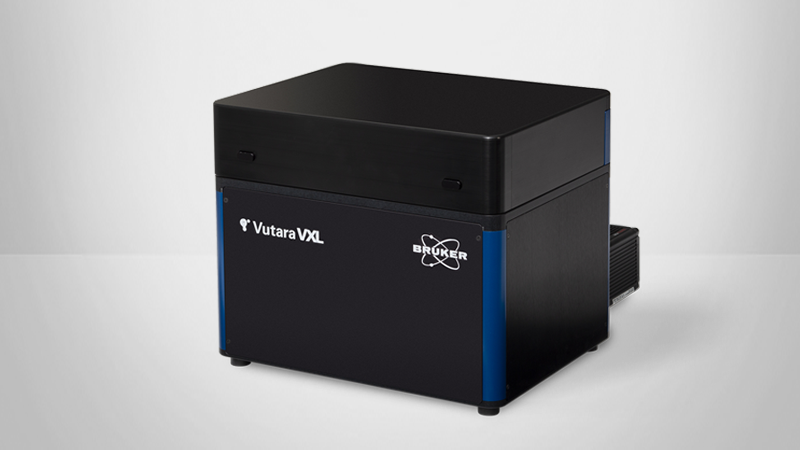
Advances in Dye Development and Microscopy for Live Cell Super Resolution Microscopy with the Vutara 352
Discover successful approaches for live single-molecule localization microscopy
Bruker experts come together to discuss different approaches for using super-resolution microscopy in your lab including:
- Fundamentals of single-molecule localization and the technological components of the Vutara 352.
- Developing dyes that spontaneously blink under physiological conditions and low laser power.
- The Vutara SRX software for analyzing live cell single molecule data.
Webinar Summary
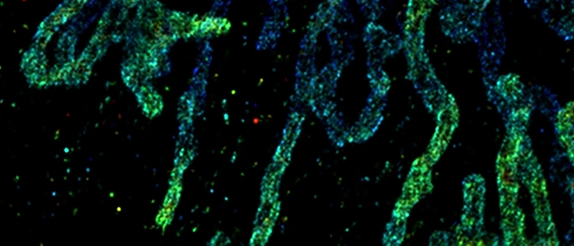
Join us for this webinar where we discuss the Vutara 352 technology and sample preparation techniques for successful live-cell imaging using currently available fluorophores. We will also describe a collaboration with Luke Lavis and Fadi Jradi (HHMI, Janelia) for developing dyes with characteristics that are ideal for long-term live-cell imaging without phototoxicity. Lastly, learn about the Vutara SRX software and its intuitive setup, calibration, imaging, processing, and analysis of data from single-molecule localization experiments.
Find out more about the technology featured in this webinar or our other solutions for Live Cell Super Resolution Microscopy:
Featured Products and Technology

Speaker

Robert J. Hobson, Ph.D.
Vutara Applications Scientist, Bruker
Dr. Robert Hobson graduated from the University of Salford, UK with a degree in Biology, before completing his PhD in Biology at the University of Toledo, Ohio. He was a research assistant professor at the University of Utah with multiple years’ experience in single-molecule localization microscopy sample preparation and imaging before joining Bruker Nano Surfaces.