
Episode 2: Applications of BioAFM in Life Sciences
Episode 2: Applications of BioAFM in Life Sciences
In episode 2 of our Conversations on AFM podcast we host Dr. Andre Körnig, an applications scientist at Bruker BioAFM. We explore the applications of BioAFM in life sciences, highlighting its versatility and ability to image biological sample's without extensive preparation.
Dr. Körnig shares his journey into AFM, emphasizing its unique ability to bridge physics and biology. Such as when studying mechanosensation in cancer research and multiparametric imaging of fibroblasts.
TO LEARN MORE:
Exploring the World of BioAFM: Why AFM is Essential in Biological Research Today
In episode 1 of our AFM podcast “Conversations on AFM”, we learned more about the origins and impact of AFM from Prof Dr Mervyn Miles. Moving on from this, we want to learn more about “Applications of BioAFM in the Life Sciences”. For this, we spoke in episode 2 with Dr. Andre Körnig, an Application Scientist at Bruker, who brings a wealth of experience in the biophysical applications of AFM.
Here, we will share what we learned from our Conversations on AFM interviews about BioAFM so far.

BioAFM is Tailored to the Life Sciences
BioAFM is tailored to analysing biological samples like cells, DNA, and proteins. Not only does BioAFM enable AFM on non-conducting samples, but it also allows the visualization of imaging of biological samples without complex sample preparation.
“AFM excites scientists and researchers alike because it combines the precision of physics with the complexity of biology.”
The minimum sample preparation necessary for BioAFM makes it suitable for imaging samples in their native environment, such as in a medium, or at a specified temperature.
BioAFM Applications over Time
BioAFM technology has developed significantly in the last few decades, and Dr. Andre Körnig notes three main changes he has observed.
1. Imaging Speed
In the last decades, microscope components have significantly improved to enable faster imaging without compromising on the image quality [1]. This means that data can now be acquired within a few milliseconds. To push this boundary further, Bruker has developed the high-speed AFM technology that increases speed even further. This makes fast scanning and screening, as well as capturing dynamics, possible.
2. Ease of Use
As BioAFM has become more widely used, the ease of use of systems has also changed. With multiple commercial systems available, microscopes specifically designed for certain applications have been developed, such as speed or multi-modal imaging. Thus, scientists no longer need to understand the ins and outs of a system but can focus on acquiring results.
3. Multi-Parametric BioAFM Imaging
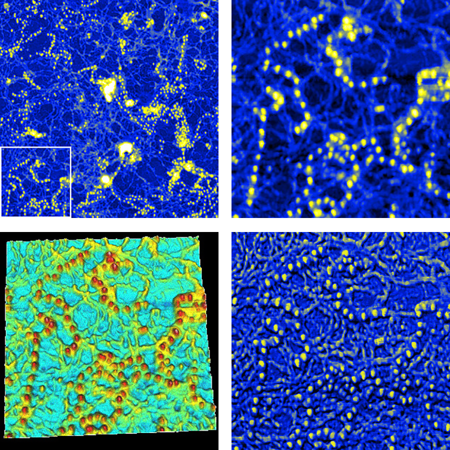
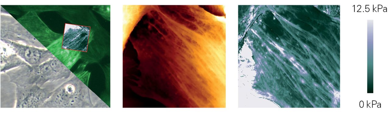
Multi-parametric imaging in BioAFM allows imaging beyond the traditional topography focus, enabling comprehensive cellular and tissue mechanics analysis [2]. Multi-parametric imaging combines data on physical characteristics like shape, stiffness, adhesion, and elasticity, which are essential for understanding biological processes and disease mechanisms. Bruker’s Quantitative Imaging (QI) mode allows researchers to study complex, soft, and sticky samples such as living cells and whole tissues without the risk of sample damage that traditional methods may cause. The Bruker QI mode enables in-depth exploration of mechanical properties, where physical characteristics play key roles in cell behaviour and disease pathology.
BioAFM Case Study: Multi-Parametric Imaging in Fibroblasts
Fibroblasts are cells central to tissue repair and inflammation. Multi-parametric imaging enables high-specificity analysis of fibroblast structure and mechanics. This can offer insights into pathologies such as fibrosis, where connective tissue scars, and fibroblast rigidity correlates with disease progression [3].
Researchers can calculate Young's modulus by recording force curves at each pixel and using contact mechanics models, revealing material properties like stress and strain. This technique also differentiates mechanical behaviour differences across the cell, with structures such as cytoskeletal F-actin and the cell membrane being visible at distinct forces. Measurements such as these provide invaluable data for understanding fibroblast mechanics and their role in disease.
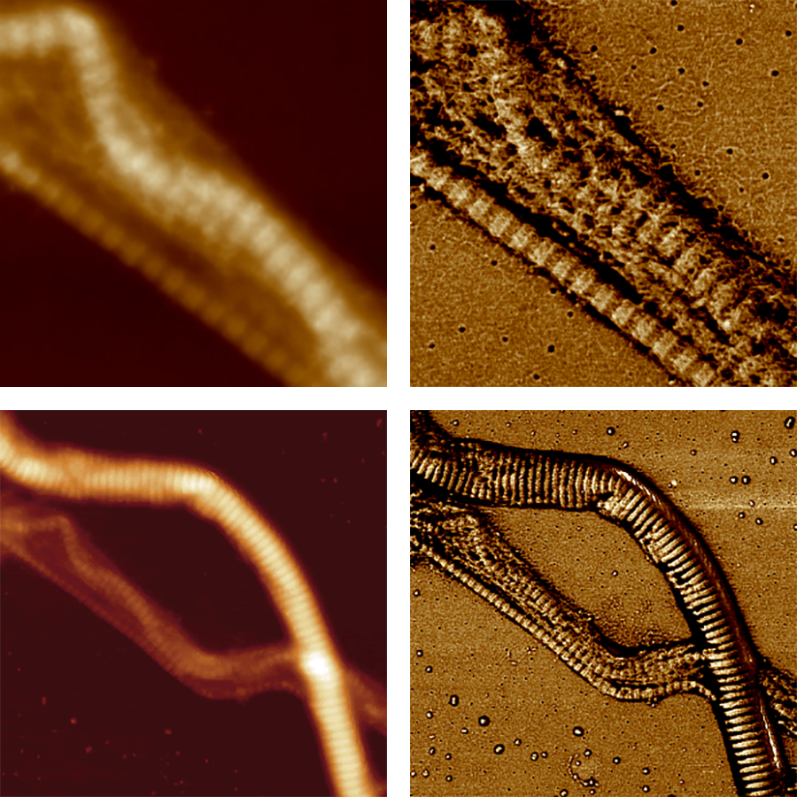
The Bruker PeakForce Tapping Module
PeakForce Tapping Mode is an AFM mode, where the cantilever tip gently taps the surface, making brief contact at very small forces. It measures the tip's maximum force on the surface (the "peak force") during each tap. This mode is especially useful for studying delicate or soft materials, like living cells, because it minimises damage while providing accurate data about their mechanical properties [4].
Dr. Körnig shared with us that the Bruker PeakForce Tapping Module is ideal for preserving probe quality and can be applied to the widest range of samples [5].
Challenges in BioAFM
As with every imaging technique, BioAFM also has to overcome certain challenges, such as that living samples can grow, move , and change over time during the image acquisition.
Additionally, due to the geometry of the setup, in BioAFM the cantilever that touches the surface of the sample comes from above. Meaning that the surface of the sample of interest has to be accessible from above.
Our Bruker application specialists, bring vast experience in sample preparation and imaging, which can help you design the ideal AFM experiment.
Conclusion
In this blog of Conversations on AFM, we have explored how BioAFM is transforming research in life sciences. Dr. Körnig shared invaluable insights into how BioAFM is used to probe cellular rigidity, investigate cell mechanics, and study fibroblast structure. His discussion highlighted BioAFM’s unique role in life sciences, offering a window into cellular processes that would otherwise remain hidden.
You can also download our ressource collection to learn more about AFM technologies and their applications.
References
- M. R. P. Ragazzon, “Parameter Estimation in Atomic Force Microscopy: Nanomechanical Properties and High-speed Demodulation,” Doctoral thesis, NTNU, 2018. http://hdl.handle.net/11250/2563513
- J. K. R. Modigunta, S. Vadivel, G. Murali, I. In, and M. Sawangphruk, “Atomic Force Microscopy: An Advanced Imaging Technique—From Molecules to Morphologies,” in Microscopic Techniques for the Non-Expert, S.-K. Kamaraj, A. Thirumurugan, S. S. Dhanabalan, and S. A. Hevia, Eds., Cham: Springer International Publishing, 2022, pp. 115–136. doi: 10.1007/978-3-030-99542-3_5.
- P. K. Viji Babu, C. Rianna, U. Mirastschijski, and M. Radmacher, “Nano-mechanical mapping of interdependent cell and ECM mechanics by AFM force spectroscopy,” Sci Rep, vol. 9, no. 1, p. 12317, Aug. 2019, doi: 10.1038/s41598-019-48566-7.
- K. Xu et al., “Recent development of PeakForce Tapping mode atomic force microscopy and its applications on nanoscience,” Nanotechnology Reviews, vol. 7, no. 6, pp. 605–621, Dec. 2018, doi: 10.1515/ntrev-2018-0086.
- PeakForce Tapping Module. https://www.bruker.com/en/products-and-solutions/microscopes/bioafm/bioafm-accessories/peakforce-tapping-software-module.html
Interested in learning more about this topic?
You can find detailed applications and technical notes, expert-led webinars, and on-demand instrument and measurement demonstrations in our online resource library. Get instant, full-length access to all resources related to this podcast using the form below.
This resource collection includes:
- 1 full-length e-book
- 2 full-length Research Highlights
- 1 full-length on-demand webinar recording
- 1 full-lenght instrument demonstration


