
Studying Morphogenetic Waves with Photo Manipulation Coupled to Multi-View Light-Sheet Microscopy
No more 2D modeling of 3D systems — learn about the latest research unraveling the mechanisms of morphogenic processes.
Watch this webinar recording to hear guest speaker Matteo Rauzi, Ph.D. (University Côte d'Azur), discusses his lab's work using a novel computational model based on light-sheet imaging, photo-manipulation, and multidimensional image analysis. This approach:
- Overcomes the limits of traditional 2D embryo imaging approaches;
- Has enabled discoveries about epithelial furrowing that challenge previous ideas about the role it plays in embryo development and the molecular signals and mechanical forces responsible for it; and
- Creates new avenues for exploring vital processes like embryo gastrulation, neurulation, and the shaping of the animal body.
Webinar Summary
Biological morphogenesis builds living shapes during embryonic development and is controlled by molecular signals and mechanical forces. While key molecular players, signaling pathways, and cellular mechanics have been studied, it is still unclear how these work at larger spatial scales.
During this 60-minute presentation, our guest speaks about their lab’s goal of uncovering the fundamental principles underlying the morphogenesis and mechanics of epithelial tissues.
Presenter's Abstract
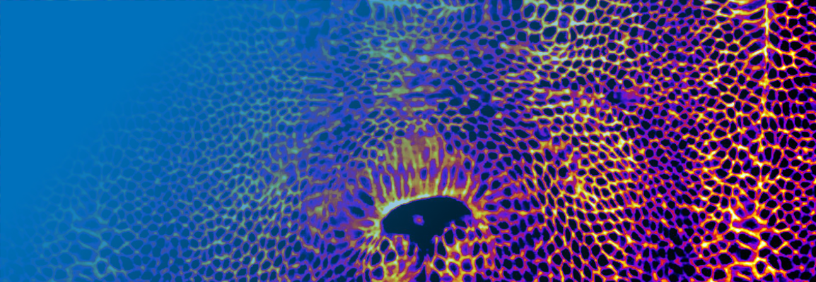
Epithelial furrowing is a morphogenetic process that is pivotal during embryo gastrulation, neurulation and the shaping of the animal body. A furrow often results from a fold that propagates along a line. How fold formation and propagation are initiated, driven and controlled is still poorly understood. To shed new light on this fundamental morphogenetic process, we study the formation of the cephalic furrow: a fold that runs along the dorsal-ventral axis of the embryo during early Drosophila gastrulation and the developmental role of which is still unknown. By implementing multi-view light sheet microscopy coupled to ultrashort infrared laser pulses manipulation and two-photon optogenetics, we provide evidence of its function and show that the cephalic furrow is initiated by two groups of cells located on the left and right lateral sides of the embryo. These cellular clusters work as a pacemaker triggering a bi-directional morphogenetic wave powered by actomyosin contractions and sustained by de novo medial apex-to-apex cell adhesion. The Cartesian position of the pacemakers is under the cross-control of the embryo anterior-posterior and dorsal-ventral gene patterning systems. Thus, furrow initiation and propagation are driven by a mechanical trigger wave that travels under the control of a multidimensional genetic guide.
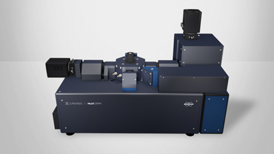
Find out more about the technology featured in this webinar or our other solutions for multi-view light sheet microscopy:
Featured Products and Technology
Guest Speaker

Matteo Rauzi, Ph.D., Group Leader at the University Côte d’Azur, Nice, France
Matteo Rauzi obtained a PhD in developmental biology, biophysics and microscopy in 2010 at the University Aix-Marseille, France. He then joined the Leptin lab as a Postdoc at the EMBL Heidelberg, Germany supported by the EMBO-Marie Curie Fellowship and the HFSP Long Term Fellowship. Since 2016, he was appointed as group leader at the University Côte d’Azur, Nice, France. Matteo is actively collaborating with Luxendo that has supported one of his PhD students.