Since cell death is very common in diseases of the brain the process of corpse removal needs to be quick and effective to prevent the manifestation of illness. In mammalian brains, microglia are regarded as the primary phagocyte responsible for corpse removal, however, under certain conditions, astrocytes have been shown to demonstrate phagocytic capability. Interestingly, whether astrocytes and microglia act in concert or independently to remove dying cells or whether each cell population has specialized functions is not known. Understanding this process might yield insights on how to address cell death in a brain injured by head trauma, stroke, and other conditions.
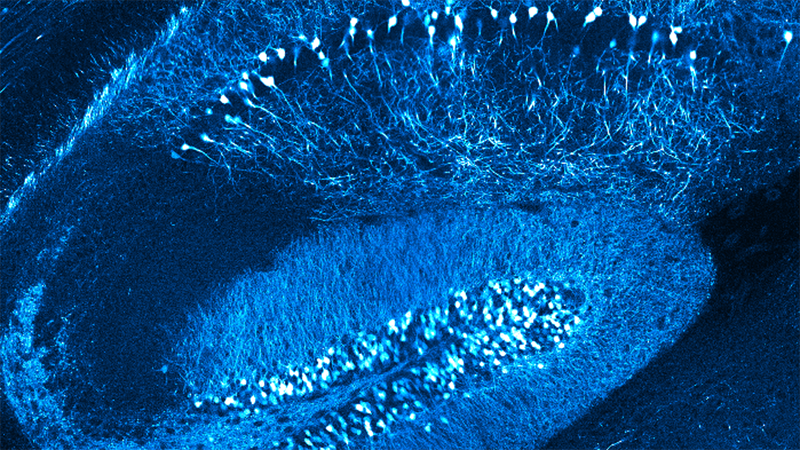
Despite the large number of cells that undergo apoptosis daily, visualization of the cell death process, the specific role of phagocytes, and the mechanisms of corpse removal in vivo have all been limited. The authors developed photochemical and viral methodologies to induce death in single cells in a live brain and combined these approaches with intravital optical imaging of the multicellular glial reaction associated with cell death and corpse clearance.
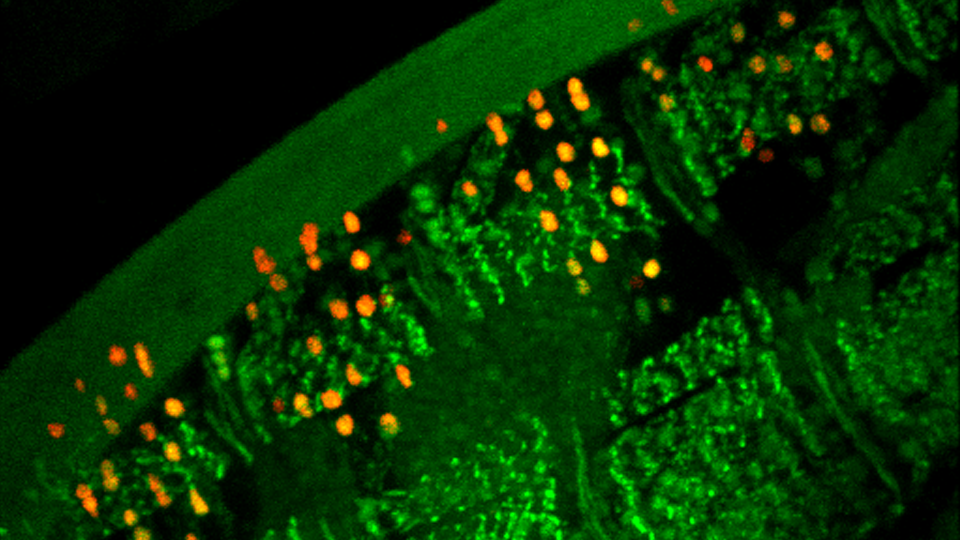
It was found that astrocytes and microglia acted in a highly coordinated fashion even though each cell type exhibited specialized function. Astrocytes surrounded apoptotic bodies derived from the extensive dendritic arbors of dying neurons by polarizing their distal processes without exhibiting cell body migration. In contrast, microglia predominantly phagocytosed dendrites, cell bodies, and nuclei by migrating toward these structures and completely engulfing them. Advanced aging was also found to be associated with a delay in removal of dying neurons.
To induce apoptosis of individual cells at a desired time and location, the 2Phatal method was used, which involved using two-photon-mediated photochemically induced apoptosis. A femtosecond-pulsed laser was used to bleach a nucleic acid binding dye within experimentally targeted cells, which induced a stereotyped apoptotic cell death process without triggering injury-related glial activation.
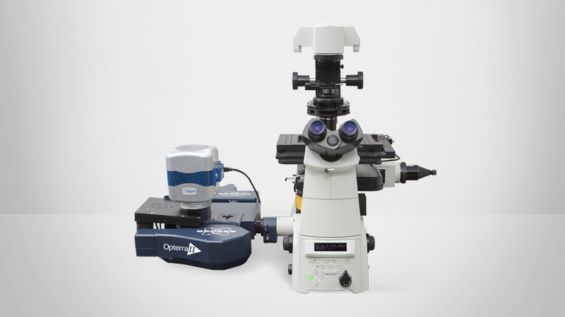
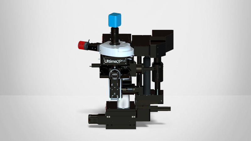
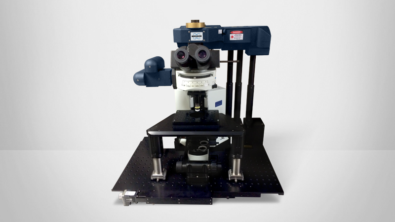
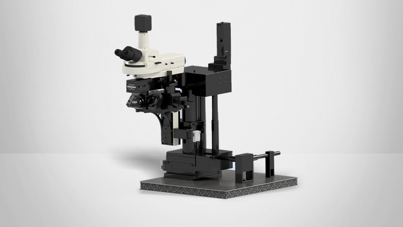
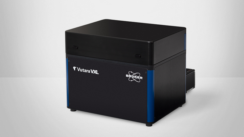
The imaging of fluorescently labeled cells in the brain of anesthetized mice was conducted using a two-photon microscope (Bruker).