LCS SPIM
LCS SPIM
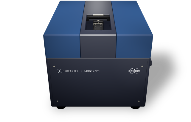
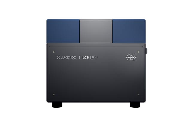
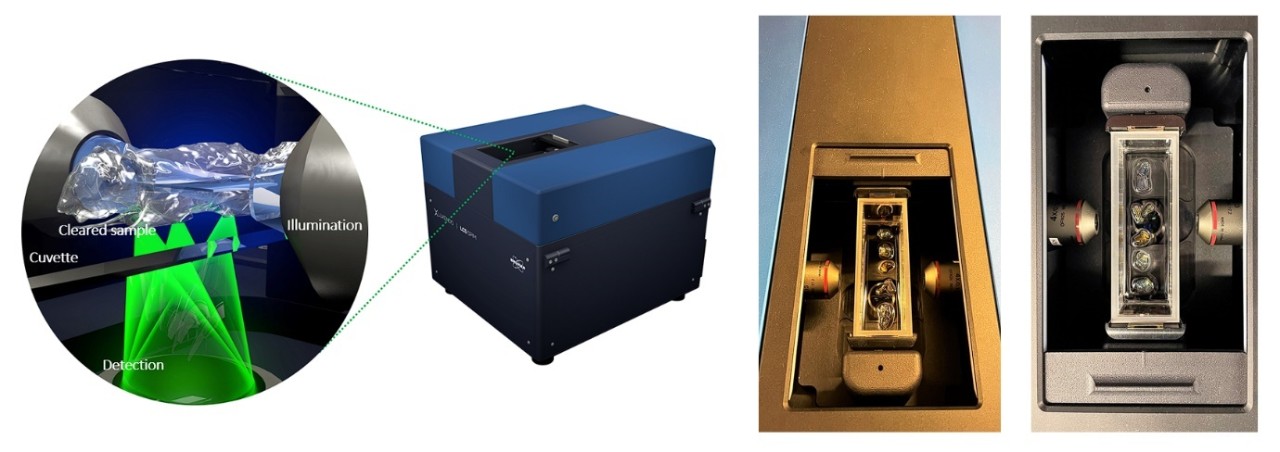
The Bruker Luxendo LCS SPIM is a compact light-sheet microscope specifically developed for large optically cleared samples. Designed with feedback from users in the lab, the LCS SPIM is easy to clean and maintain to ensure safe handling of clearing liquids. Importantly, the LCS SPIM is compatible with all currently available clearing solutions which makes it an optimal system for multi-user environments and experiments using different clearing approaches. Flexible embedding options also support small and large samples. Overall, an inverted optical concept, three objective lenses, dual-side illumination, and single-side detection ensure low sample distortion, a fast-imaging option, and an extended field-of-view (FOV). The LCS SPIM is the ideal microscope for high-throughput studies and multi-user environments.
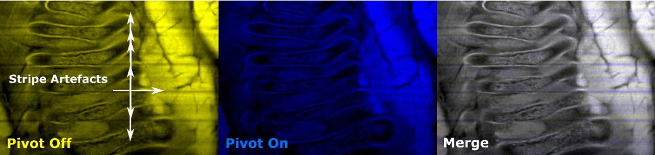
Dual-Destriping: Impressive Elimination of Striping Artifacts
When imaging, obstacles like pigments or cell nuclei can interfere with light, leading to images with shadows and striping artifacts. The next-generation SPIM employs a dual de-striping via pivot scanning and beam swiping, which generates a homogenous illumination profile without compromising acquisition speed.
TAG-Lens: Light-Sheet Length Control with Axial Scanning
Thanks to state-of-the-art technologies, ultra-fast varifocal lenses can be used to fine-tune the system's optics. Luxendo SPIMs use devices that are driven by an acoustic wave, called tuneable acoustic gradient lenses or TAG lenses. These lenses allow better light-sheet control and uniformity across the FOV.
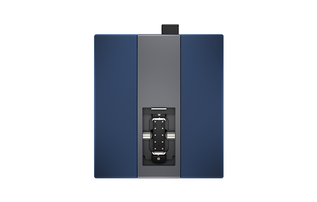
Compact, Benchtop Design
The LCS SPIM's compact, robust and vibration-free design provides maximal stability even during long-term high-throughput experiments.
The basic configuration of the LCS offers a cost-effective solution for cleared sample imaging. Based on a very versatile layout, the highly flexible upgrade options expand the performance of the system to increase speed and optical performance.
Tailored to fit your lab bench, this class 1 laser system does not require any air table or vibration-compensation mechanism as all moving components are light-weight and balanced. Maximal stability of focus and thermal conditions are also guaranteed. The proprietary piezo-crawler stages ensure longevity and precision for a permanently accurate specimen positioning. Neither the images nor the quality of your sample are affected thanks to the unique and very gentle image-acquisition concept.
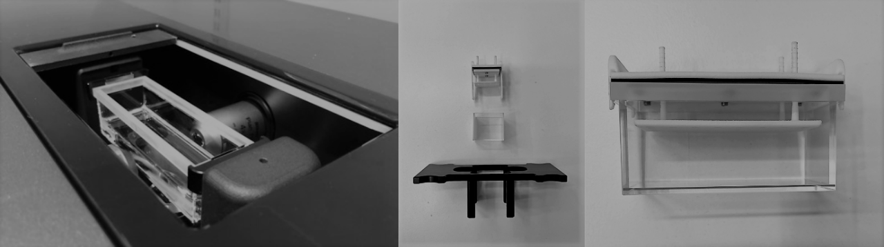
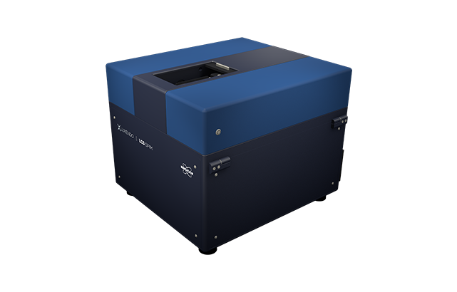
Large and Closed Cuvette
Using exchangeable quartz-crystal cuvettes simplifies sample mounting procedures as well as microscope cleaning and maintenance. Three standard cuvette sizes from [20 mm × 20 mm × 20 mm] up to [100 mm x 30 mm x 30 mm] (l × w × h) allow imaging from organs to organisms.
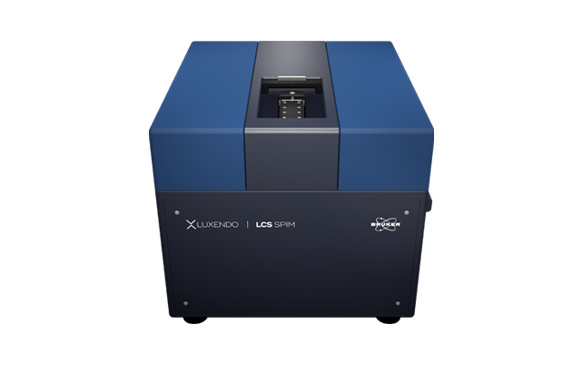
Illumination and Detection
To ensure the best illumination, the cuvette moves in z, followed by the focus shift being corrected by changes with the detection optics. The robust sCMOS camera is well-suited for experiments that require high detection efficiency, quantification, and speed.
The Ideal Setup for Cleared Samples
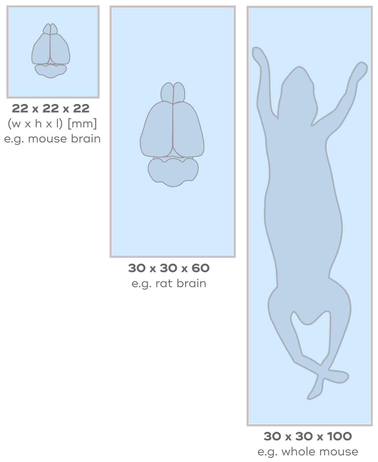
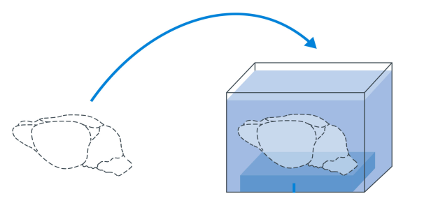
High Flexibility of Sample Size
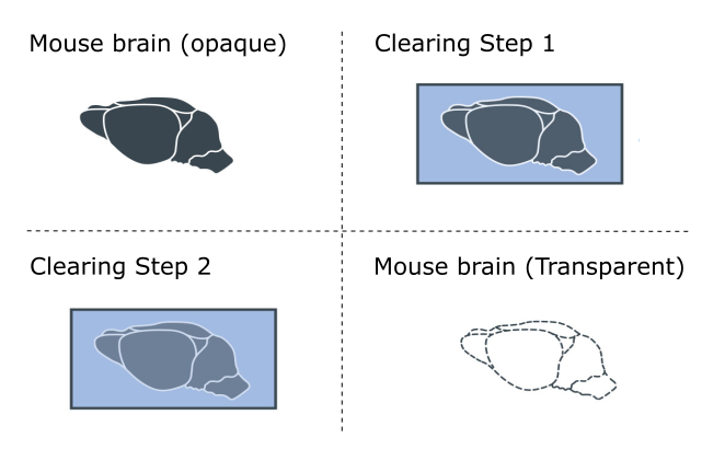
Whether it is cleared organs or whole organisms, such as from a brain to a whole mouse, the flexibility of the LCS SPIM allows a wide flexibility of imaging options. all of those to be imaged. Importantly, samples are placed into closed cuvettes, which makes sample handling safe and ensures compatibility with all clearing solutions by covering a refractive index (RI) range from 1.33 to 1.58.
Most microscope systems are incompatible with more than one clearing approach as different tissues and samples often require different clearing approaches and solutions. However, the LCS SPIM is a truly flexible system designed to be compatible with any currently available clearing solutions.
Advanced Fast-Imaging Acquisition
Using axial movement of the light-sheet extends the effective FOV of the illumination beams. This means that fewer images are needed, imaging rates are higher, and phototoxicity is reduced. Additionally, the image quality is increased by a reduction of shadowing artifacts.
Importantly, the high speed of imaging made possible with LCS SPIM and Advanced Fast-Imaging Acquisition is great for imaging large cleared samples. The LuxBundle processing software also allows for fast and accurate image processing, which is important when imaging and processing data from large cleared samples.
Example Speed Scanning a Whole Mouse Brain with 2 labels/channels:
Magnification: 4.4x - Tiles: 5x4 tiles (20 total) Resolution: 1.4 x 1.4 x 10 µm (x,y,z) – Exposure Time: 50ms – Scan Time: 30 min – Processing: 30 min
Innovative and Easy Sample Handling
Robust, safe, and reproducible sample handling is key. The LCS SPIM comes with three standard sized closed cuvettes from [20 mm × 20 mm × 20 mm] up to [100 mm x 30 mm x 30 mm] (l × w × h), allowing the mounting from small organs to medium-sized organisms. Having three sizes means that the cuvettes will fit the samples and only a minimum of the usually expensive clearing solutions is needed.
As the cuvettes cover the typically used refractive index (RI) range from 1.33 to 1.58 and more, they are compatible with all currently available clearing solutions. To allow for further customization, easy-to-use sample holders were designed together with end-users and application specialists. Furthermore, there is an option for users to use 3D printing to produce customized sample holders.
High-Throughput Capability
A quartz crystal cuvette facilitates the fast mounting of large, delicate samples. The specimen is simply introduced into the cuvette filled with the clearing solution.
The motorized sample stage for positioning and stack acquisition as well as the programmable optics concept for fast 3D scanning of the light-sheet through the sample ensure that the sample remains unperturbed during the imaging experiment while achieving the highest acquisition speed.
Illumination and Detection
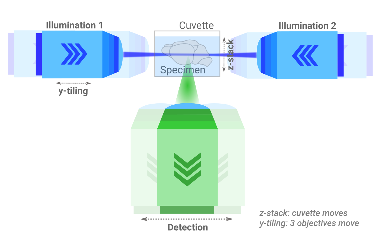
The illumination optics comprise light-sheet generation by scanning a beam after passing a beam-shaper unit for length and diameter adjustment. Two Nikon 4x 0.2 NA air objective lenses project two aligned light sheets from opposing directions on the sample. The expandable and upgradable illumination setup allows a very flexible choice of lasers.
The detection optics comprises one Olympus 4x 0.28NA air/dry lens. An additional magnification changer results in a total magnification between 2.2x and 8.8x. The spatial detection arm is equipped with a 10 position high-speed filter wheel. The robust sCMOS camera is well-suited for experiments that require high detection efficiency, quantification and speed.
LSC SPIM Design
WHOLE ORGAN IMAGING - GEOMETRIC PATTERNING OF TISSUES.
The combination of tissue clearing, and light-sheet imaging is probably the most famous for creating in-depth 3D insights of whole organs and tissues. Combining tissue clearing with state-of-the-art imaging provides novel insights into the in-toto geometric patterning of tissues, organs, and organisms.
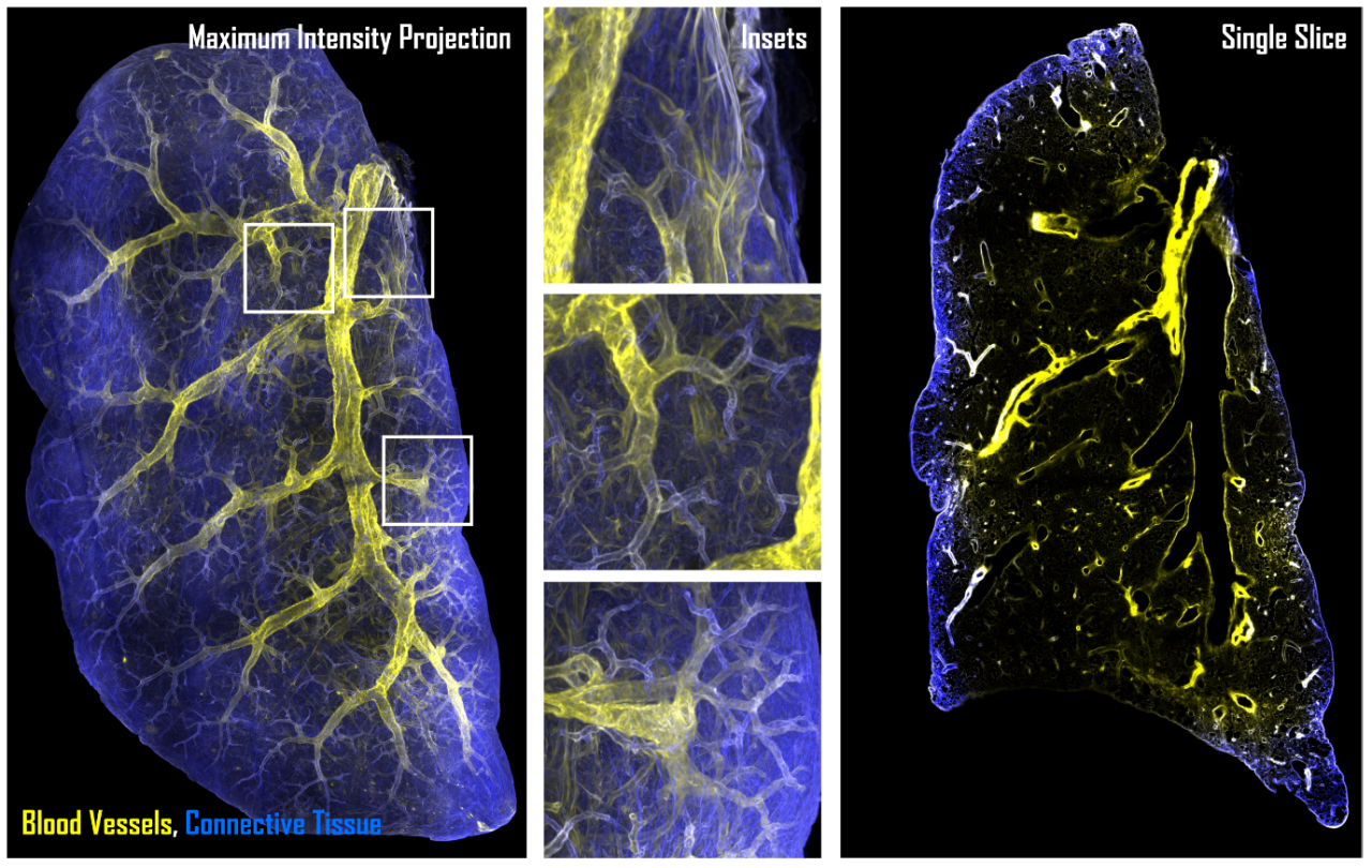
Samples courtesy of: Ayelen Melina Santamans Recchini and Gaudalupe Sabio Buzo., Stress kinases in Diabetes, Cancer and Cardiovascular Disease Laboratory and Unit of Microscopy and Dynamic Imaging, Centro Nacional de Investigaciones Cardiovasculares Carlos III (CNIC), Madrid.
MINIMAL SOLUTION REQUIREMENTS.
Using the correctly sized cuvettes for sample embedding allows for the use of minimal clearing solution, saving money and reducing chemical exposures.
WHOLE MOUSE IMAGING.
Images courtesy of: Dr. Chenchen Pan, DKFZ, Heidelberg, Germany.
NEUROGENESIS.
Understanding how the nervous system is formed and maintained is critical to perceiving how the nervous system changes with age or in disease. Using tissue clearing, combined with light-sheet imaging on the LCS SPIM provides novel 3D insights in intact organisms. Thus, studying intricate nerves in whole animals is possible and no longer limited to medical imaging modalities.
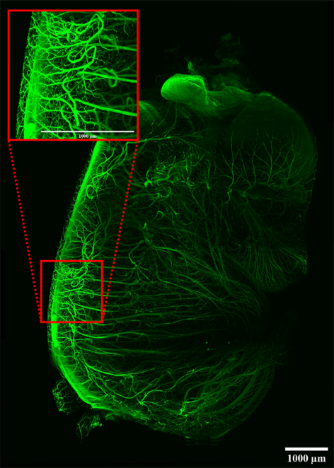
Sample cleared with DBE. Tiled image (3 x 4) acquisition. Scalebars: 1mm.
Imaged on the LCS SPIM.
Sample courtesy of: James Muller, MSKCC, New York, USA.
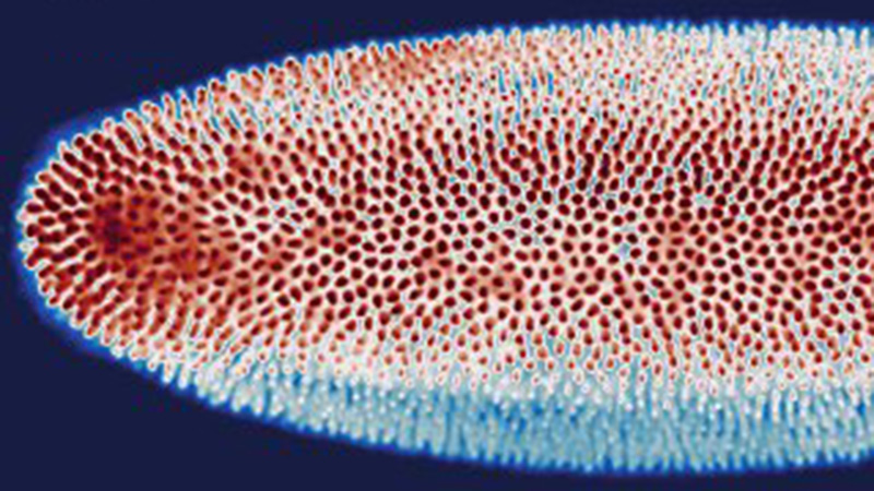
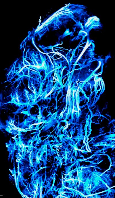
MORPHOGENESIS AND HEALTH.
Imaging with the LCS SPIM is not limited to already formed tissues but helps to understand how tissues are formed and maintained. In this example, muscle-specific precursors are visualized in a fruit fly. This helps to understand how muscle tissues form in development and are maintained in health.
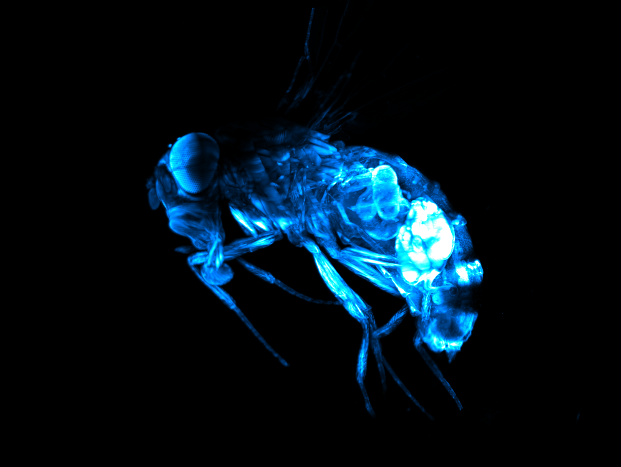
The fruit fly was optically cleared, and refractive index adjusted using alcohol dilutions and ethyl cinnamate (ECi). Length head-to-tail: 1.7 mm length.
Imaged on the LCS SPIM.
Courtesy of: Susanne Önel, Philipps University Marburg, Marburg, Germany.
Cleared-Sample / Cleared-Tissue Applications
Browse a selection of applications data from our customers below. Researchers are using LCS SPIM in a variety of ways including studies.
Specifications
| Detection | Eff. Magnification | FOV [µm] | Pixel Size [nm] | Optical Resolution |
| Olympus 4x 0.28 NA | 2.2x | 5990 | 2925 | 1.1 µm |
| 4.4x | 2995 | 1463 | ||
| 6.7x | 1997 | 975 | ||
| 8.9x | 1497 | 731 |
System Comparison
* COMPATIBLE MODULE
| The perfect allrounder - multi-view imaging for diverse samples (live and cleared option) | Multi-sample and dual view flexibility for highly sensitive imaging, ideal for organoids and 3D cell cultures | In vivo multi-sample and multi-condition imaging, ideal for drug treatment comparison | Perfection for cleared samples prepared by any type of clearing protocol | High-end imaging with various beam patterns from single cells to 3D cell cultures | |
|---|---|---|---|---|---|
| Geometry | Horizontal with multiview | Horizontal with dual view | Inverted with dual illumination | Inverted dual illumination with moving optics | Inverted geometry at 360° |
| Beam Type | Gaussian | Gaussian and Bessel | Gaussian | Gaussian | Advanced Illumination Module (AIM) |
| Application Examples | - Long-term imaging of Drosophila development - Photomanipulation studies in zebrafish embryos - Cleared and stained samples up to mouse brain size | - High-resolution full 3D imaging of multiple organoids over time - Long-term imaging of highly light-sensitive samples - Comparing the pharmacological impact in zebrafish wound healing after PM | - Time-lapse imaging of multiple pancreatic spheres - Studying pharmacological impact on zebrafish development - Comparing genetically altered organoids | - Pharmacokinetics in mouse brain - Multi-stained rat brain - Whole mouse imaging | - Fast imaging of photo-sensitive 2D cell culture - Lattice imaging for enhanced resolution in 3D cell cultures - FCS and FLIM in cell culture |
| User Level | Beginner friendly | Beginner friendly | Beginner friendly | Intermediate | Advanced |
| # Lenses | 4 lenses (2 IO, 2 DO) | 3 lenses (1 IO, 2 DO) | 3 lenses (2 IO, 1 DO) | 3 lenses (2 IO, 1 DO) | 2 lenses (1 IO, 1 DO) |
| Multi-View | ✓ | ✓ | |||
| Live/Fixed Samples | ✓ | ✓ | ✓ | ✓ | |
| Cleared Samples | ✓ | ✓ | |||
| Expanded Samples | ✓ | ✓ | ✓ | ✓ | ✓ |
| Best Embedding | Capillary/FEP tube w/ agarose 3D stage | Custom-solution and FEP foil | TruLive3D dishes and FEP foil | Quartz-crystal cuvette | FEP foil; glass slides |
| Photomanipulation* | ✓ | ✓ | ✓ | ✓ | |
| Environmental Control* | ✓ | ✓ | ✓ | ✓ | |
| Destriping/Uniform Illumination* | ✓ | ✓ | ✓ | ✓ | ✓ (w/ Advanced Illumination Module) |
| Benchtop Design | ✓ | ✓ | ✓ | ✓ | ✓ |
All-in-One LuxBundle Software
Intuitive design
Bruker's LuxBundle software saves time and enhances productivity by providing:
- All-in-one, easy-to-use interface for acquisition, viewing, and post-processing
- Fully scriptable microscope control and post-processing via open interface (e.g., Python or any other language), ready for custom "smart" microscopy
- High reproducibility of experiments: all parameters are saved in the metadata and configurations can be saved for future experiments
- Data formats (.tiff, .hdf5, .ims) compatible with common image processing software: Imaris, Aivia, BigDataViewer, Arivis, Fiji, Python, Matlab, Napari
Impressive Image Post Processor
Our dual-view light-sheet microscopes record a sample from different angles/views and generate images composed of multiple tiles. The LuxBundle software ensures high-quality, 360° crisp images of the sample that compensate for absorption and scattering. Features include:
- Multi-color alignment
- Tile stitching of hundreds of tiles for large samples
- Multi-view image fusion and deconvolution
3D Data Viewer
LuxBundle's integrated 3D data viewer allows researchers to inspect the entire dataset directly after acquisition. This gives users control over their data with key capabilities, including:
- The ability to turn tile stitching on or off
- Both raw and post-processed images
- Fast viewing of multi-terabyte data sets
- Flexible options to draw and annotate regions and landmarks