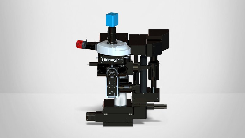
Inverter Module
Expanding Multiphoton Imaging Beyond Upright Geometry
Bruker’s new Inverter Module for Ultima 2Pplus and Investigator Plus multiphoton microscopes enables inverted two-photon imaging without sacrificing optical performance. The Inverter Module quickly and easily transforms Bruker’s upright multiphoton platforms into a flexible inverted configuration, opening the door to high-quality imaging of slides, dishes, organoids, and plated cells while preserving the strengths of the original upright system.
The Inverter Module was developed to address the need to efficiently image samples that settle or adhere to the bottom of dishes and plates. Using custom relay optics and a mirror-based light-routing assembly, the module redirects the excitation and detection paths so that the objective lens points upward, creating a true inverted configuration. The result is a single system that supports both upright and inverted imaging modalities, expanding experimental flexibility and reducing the need for separate microscopes.

How the Inverter Module Extends Ultima System Capabilities
In this video: Jimmy Fong (Director, Products and Technology, Bruker Fluorescence Microscopy) provides an overview of the Inverter Module, explaining how it extends Ultima 2Pplus and Ultima Investigator systems to effectively function as two microscopes in one. He walks through what the module is, how its design was driven by real research needs, and the ways it expands experimental flexibility without the need for a separate standalone setup.
The overview also highlights example experimental data and the types of applications and experiments the module makes possible.
Contact us or continue reading to learn more.
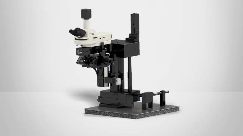
True Upright‑to‑Inverted Conversion
Complete optical reorientation
The Inverter Module enables a true upright‑to‑inverted conversion by fully reorienting the system’s optical geometry. Its custom relay lens and mirror‑based light‑routing assembly redirects the excitation and detection paths so the objective points upward instead of downward, placing the sample above the objective — the correct geometry for slides, dishes, multi‑well plates, and plated tissues.
Preserved optical performance
The Inverter Module was designed to maintain the performance of Bruker multiphoton systems, avoiding the light loss and reduced image quality of workarounds and partial modifications. Its custom optics and mechanics maintain full signal throughput, optical alignment, and image fidelity, enabling inverted imaging with no trade-offs.
No second system required
The Inverter Module turns a single system into a 2‑in‑1 platform for both upright multiphoton and inverted imaging. Dish‑based assays, organoids, plated cells, and tissue samples can be imaged on the same instrument, for both in vivo and ex vivo neuroscience studies. This eliminates the need for a separate inverted microscope.

Purpose‑Built for for Safe, Simple, Real‑World Operation
Seamless on-site integration
The Inverter Module was designed to integrate directly into Bruker multiphoton systems with fully on-site installation. Installation can often be completed in a day, minimizing research disruption and allowing labs to expand capabilities quickly.
Intuitive dual‑mode operation
Once installed, the module lets users switch easily between upright multiphoton imaging and inverted workflows on their own. Because it works with Bruker's standard XY stage and preserves system capabilities, researchers maintain familiar motion control and sample‑handling workflows while gaining access to inverted formats such as slides, dishes, plated tissues, and multi‑well plates.
Safe for inverted configurations
To enable safe operation in an inverted geometry, a light‑tight, safety‑interlocked chamber prevents exposure to upward-facing laser beams. The laser can operate only when the enclosure is closed, ensuring safe use in both individual labs and shared facilities.


Examples from Current Research
Imaging of Neural Circuits in Live Spheroids
The following two examples illustrate how the Inverter Module supports organoid workflows in both dish‑based and in vivo contexts.
In Image 1: Organoids grown and plated in dishes were imaged using the Inverter Module, allowing the lab to perform high-quality two-photon imaging in an inverted configuration optimized for dish-based samples.
In Image 2: Neural circuits developed in a live spheroid, with organoids grown and validated ex vivo in an inverted configuration prior to implantation for upright in vivo multiphoton imaging. The inverter module supports this integrated workflow on a single system, enabling easy progression from dish‑based validation to in vivo imaging and supporting studies of transplantation, integration, and regeneration.


Inverted Imaging of Cancer Cells Reveals Metabolic Insights
Researchers have applied the Inverter Module to image plated cancer tissue samples, including multicolor and autofluorescence imaging. Large tissue areas can be acquired using XY stage scanning and stitched into high-resolution montages, supporting metabolic and structural analysis.
Image 1: Fresh, human‑derived cancer tissue was plated on a dish and imaged using the Inverter Module, capturing GFP and native autofluorescence signals in an inverted setup. The workflow produced high‑quality two‑photon images of real tissue samples in standard dish‑based formats.
Image 2: A larger cancer‑tissue sample was imaged through a stitched‑field acquisition, using the XY stage to collect multiple adjacent fields of view and merge them into a continuous multicolor image. This approach enabled large‑area, plate‑based tissue imaging suitable for examining tumor structure and metabolism.


Frequently Asked Questions
Capabilities
It supports both upright multiphoton imaging and inverted workflows on the same system.
It supports slides, dishes, multi‑well plates, plated tissues, and organoids — formats typically associated with inverted systems — while retaining upright capabilities for animal and tissue imaging.
By enabling both upright and inverted imaging on one multiphoton system, researchers can move from dish‑based validation to in vivo animal imaging without switching instruments. This dual‑use capability expands support for non‑neuro applications while still accommodating traditional in vivo and ex vivo neuroscience workflows.
Usability
The inverter module retains the 14 degree high collection angle of the upright configuration as well as the field of view capabilities.
Once installed, users can switch between upright multiphoton imaging and inverted modes quickly and easily on their own.
The module is aligned in the factory and checked at installation such that switching configurations does not affect the alignment. Calibrations are set during the installation so that they can be loaded in software.
Usage of the inverter arm will be trained during installation including sample mounting and example imaging protocols. Both upright and inverted configurations are driven by Prairie View software. Please contact your Bruker FM representative for any assistance in updating software.
Yes. The module increases flexibility for shared labs by supporting both animal and non‑animal workflows and enabling higher instrument utilization across diverse user groups.
Compatibility and Integration
The Inverter Module is compatible with Ultima 2P Plus and Ultima Investigator systems.
Yes. The module is field‑upgradeable and can be installed on existing systems without sending the microscope back to the factory.
The module works with Bruker’s standard XY stages and supports slides, dishes, multi‑well plates, and plated tissues.
The module is compatible with the objective configurations already supported by the Ultima 2Pplus and Ultima Investigator systems; no special objectives are required.
Yes, the inverter module with stage follows sizing conventions of other inverted microscope stages and are compatible with environmental chambers from many manufacturers.
The inverter does not change the working distance achievable with a given objective. Also, the module fits within the footprint of a lightbox enclosure for the 2Pplus and Investigator Plus microscopes.
Installation and Other Technical Considerations
Like our other multiphoton microscope modules, the Inverter Module is available as a separate add‑on rather than a standard inclusion.
Yes. It is designed for on‑site installation, which typically takes about a day and does not require major system disruption.
No. Users cannot install the Inverter Module themselves. A Bruker expert performs the on‑site installation—typically completed within a day—and ensures the system is configured correctly. After installation, users can operate the module independently and switch between upright and inverted configurations on their own, without technician assistance.
No. The module is fully field‑upgradeable, and systems do not need to be returned to the factory for installation.
For additional information about the Inverter Module, you can download the datasheet or contact a Bruker expert directly. You can also request a quote or pricing information via our online quote request form.