
Observing Viral Structure and Host-Virus Interactions
SMLM for powerful virology insights
Attendees will gain first-hand insights into how SMLM surpasses the diffraction limit for viral research. Applications made possible with this super-resolution technique include:
- Understanding virion structure
- Monitoring virus-cell interaction
- Viral in situ genome imaging
- Live cell and particle tracking
Webinar Summary
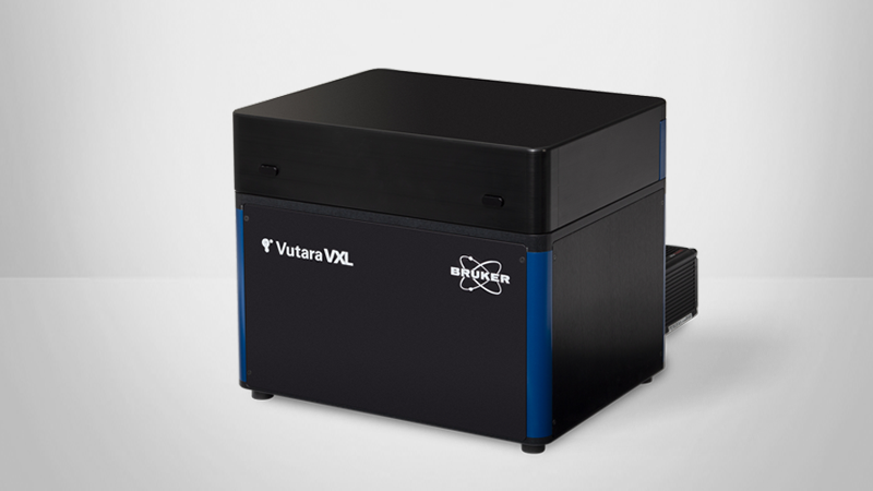
Single-molecule localization microscopy provides the ability to image biological processes well below the diffraction limit of light. Viral particles are often less than 300 nm in size, leaving super-resolution microscopy as the only fluorescence technique that can observe viral structure or host-virus interactions in high spatial detail. This webinar will highlight the use of the Vutara 352 super-resolution microscope in virus research. We will cover the basics of single molecule localization microscopy and highlight research papers that use the Vutara to answer questions about virus biology: from the structure of individual viral particles to the interactions of viral particles and components with host cell proteins in situ. Finally, we will show how the Vutara could be used in live cell applications and genome imaging for virus research.
Find out more about our other solutions for Super-Resolution Microscopy:
Featured Products and Solutions
Speakers
Robert J. Hobson, Ph.D.
Vutara Applications Scientist, Bruker
Dr. Robert Hobson graduated from the University of Salford, UK with a degree in Biology, before completing his PhD in Biology at the University of Toledo, Ohio. He was a research assistant professor at the University of Utah with multiple years’ experience in single-molecule localization microscopy sample preparation and imaging before joining Bruker Nano Surfaces.

