Experiences in the Use of Micro-CT in Insect Research
Webinar Overview
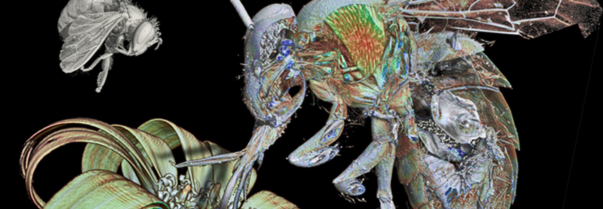
High resolution microtomography (micro-CT) despite it is not a new technique, nowadays it is being used routinely in science. Mostly it is being used as a substitute to microscopy to look at/for external and internal structures. However, it can be used to clarify non-answered questions, and also for educational purposes. And in many cases simply it represents itself, Art.
The main advantage is that it is a non-destructive technique, and once the sample has been scanned, the reconstructed images can be stored, and at any time viewed, rotated, sliced, etc…, making it possible to study details of external and internal structure in any possible perspective. The author uses a high resolution Skyscan desktop micro-CT with a maximun voxel size resolution 0.48µm. He will share experiences of different studies exemplifying how micro-CT resulted an unvaluable tool to unveil functional anatomical, as well biological aspects of different insect orders and species. The talk will be illustrated with high resolution images, and videos to demonstrate how thanks to micro-CT it is possible to do amazing trips inside the insects. Moreover “real” entomological models can be visualized, rotated, cut, etc…, in an interactive mode, when using mobile devices (smartphones and tablets), resulting good tools for research, very attractive for the students, and thereafter very promising for educational purposes. In any case, many of the obtained results have a nice visual artistic appearance, and attenders will understand why we consider that the Micro-CT represents a tool straddling scientist research, art and education.
Thursday June 17, 2021
04:00 PM CEST
Key Topics
- Use of microCT for anatomical research in insects and other species
- Contrasting techniques for microCT imaging
- 3D visualisation of microCT data
- Advantages of microCT compared to histology for anatomical analysis
Who Should Attend?
Academic researchers, doctoral students, post-docs, biologists, biomedical scientists and museum scientists in the field of insect and zoological research an classification biology.