Application Note: Behavior-Based, Closed-Loop In Vivo Optogenetics and Imaging with nVoke and nVision
Seamlessly integrate real-time behavior tracking, calcium imaging, and optogenetics
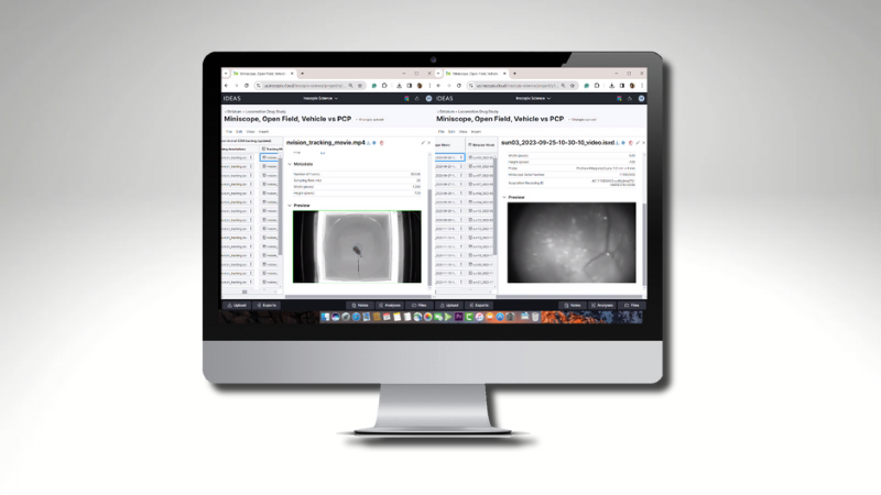
In this application note, readers will learn about how the Inscopix nVoke and nVision systems are used in freely moving animals to condition real-time place preference. Read about data examples analyzed in the IDEAS platform, highlighting our seamlessly integrated end-to-end workflow for complex behavioral neuroscience experiments.
Readers can expect to learn more about:
- nVision paired with nVoke enables real-time behavior tracking, calcium imaging, and optogenetic stimulation in freely behaving mice
- Real-time place preference (RTPP) experiments reveal activity of a distinct subset of cells
- Validated protocols using ChrimsonR and GCaMP8, including stimulation parameters and behavioral zone assignments
- The combination of nVision, nVoke, and the IDEAS cloud-based analysis platform offers a seamless workflow, making closed-loop experiments easier to conduct and analyze
KEYWORDS: nVision, nVoke, miniscope, IDEAS software, neuroscience, brain imaging, optogenetics

Introduction
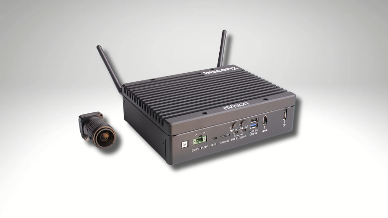
The optical toolkit available to neuroscientists in recent years permits the simultaneous manipulation and observation of multiple cell populations along a neural circuit. Miniscope technology allows for free behavior during imaging and optogenetic stimulation, granting researchers unique insights into the consequences of circuit activation on naturalistic behavior or in the context of learning, memory, and reward. Closed-loop manipulations that couple behavior to direct activation of these circuits are extremely powerful tools for understanding the neurobehavioral landscape of motivated behaviors, but the technical challenges of online behavior tracking have limited the extent of these investigations. The nVision™ closed-loop application offers seamless integration of real-time behavior tracking, calcium imaging, and optogenetics. Paired with the IDEAS data processing and analysis platform, the nVision closed-loop application offers a simplified end-to-end workflow for rapid data collection and processing of complex behavior-informed optogenetics experiments using the nVoke™ system (Figure 1).

Behavior-based in-vivo optogenetics and imaging
Leveraging the nVision closed-loop application and the nVoke opto-genetic imaging system, we demonstrate the ability to condition a real-time place preference using zone occupancy-based cues to inform optogenetic stimulation of a known reward circuit while simultaneously imaging GCaMP activity in the nucleus accumbens.

Materials and supplies
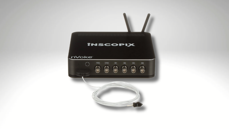
The nVoke system consists of a miniature microscope, data acquisition box, Inscopix Data Acquisition and Processing Software (IDAS) including direct upload capability to our cloud-based IDEAS data processing solution, and hardware accessories (Figure 3). The nVision system includes behavioral cameras, hardware accessories, and IDAS that permits pairing to nVoke, nVue, and nVista systems for time-locked neural and behavioral recordings, as well as live animal tracking and closed loop functionality to inform complex miniscope protocols based on animal behavior.

Experimental Procedure
A viral construct encoding the red-light driven opsin Chrimson was injected into the left basolateral amygdala of 8-10 week old male C57BL6/J mice using pulled glass capillaries. 2 weeks later, jGCaMP8m was injected into the nucleus accumbens and a 0.6x7.3 ProView integrated lens was implanted in the same region dorsal to the injection site.
Behavior Protocol
Three weeks after lens implantation, mice were mounted with the nVoke miniscope and placed into an open field arena with memory markings on one side. Zones were defined within the arena and assigned to triggers such that occupancy of one zone but not the other would initiate simultaneous calcium imaging and engagement of an optogenetic pulse protocol (5 ms pulse width, 20 Hz pulse frequency, 2 second pulse train duration, 2 second inter-train interval) for the duration of occupancy. Miniscope recording continued with no optogenetic stimulation when the mouse crossed into the opposite zone not assigned to an OG-LED trigger. For Real-Time Place Preference (RTPP) experiments, mice were first habituated to the weight of the miniscope in home cages. Following habituation, all mice underwent a 30-minute pretest session consisting of continuous Ca2+ imaging without OG-LED activation in order to assess initial preference. The arena was divided into zones designated as either "stripe" or "blank," based on memory markings placed within the arena, and behavior was recorded to determine pre-test zone occupancy. Two subsequent days of RTPP testing followed. The arena zone designations remained the same as during the pretest sessions, with OG-LED triggers now assigned to each zone such that entry into whichever zone was least preferred by the animal during the pretest phase was paired with the OG-LED pulse protocol (OG-LED+ EX-LED), with no OG-LED trigger assigned to the opposite zone (EX-LED only).
RTPP imaging sessions consisted of a 5-minute baseline where no OG-LED stimulation was present, followed by 30 minutes of recording with behavior-triggered OG-LED pulses assigned to occupancy of a designated zone (Figure 4C). Sessions were completed in 10 mice and counterbalanced such that opposite sides of behavioral arenas contained memory markings. Following RTPP testing, an additional 2 days of recordings were performed in the presence of OG stimulation assigned at regular intervals regardless of zone occupancy.

Results
Of the 10 animals tested, 4 showed preferences for the stim-assigned zone during RTPP sessions, relative to occupancy of the same zone in the absence of OG-LED stimulation during the pretest phase. Percentage of time spent in the stim-assigned zone was similar to that observed during pretest sessions when OG-LED stimulation was delivered in both zones at regular intervals (Figure 5B). Traces extracted from RTPP sessions displayed increased, synchronous activity during periods of time for which the OG-LED was on, and a distinct subset of cells was activated by OG-LED stimulation (Figure 5A).

Discussion
Optogenetic manipulation of neural circuits allows re-searchers to precisely control and monitor the mecha-nisms of complex behaviors, facilitating the acquisition of preferences and reward-based learning in real time (5, 6). Drawing on our previously published proof-of-concept work using closed-loop optogenetics with EthoVision to probe the BLA-to-NAc circuit (7), we’ve used the newly developed nVision closed-loop application combined with the nVoke optogenetic miniscope to carry out a real-time place preference assay while simultaneously performing GCaMP imaging in the nucleus accumbens in freely-behaving mice. Combining nVision live tracking and behavior-informed triggering of an optogenetic protocol to deliver stimulation to Chrimson-expressing cells in the basolateral amygdala, we demonstrate a robust preference for the stimulus-paired side of an open field arena. We also leverage the IDEAS cloud-based analysis platform to perform complex primary and secondary neural and behavioral analysis, offering a seamlessly integrated end-to-end workflow for the complex manipulation, observation, and analysis of cellular activity and behavior in real time.
References
- Carter, M.E., & de Lecea, L. Optogenetic investigation of neural circuits in vivo. Trends Mol Med 17(4): 197-206 (2011).
- Grove, J.C.R., et al. Dopamine subsystems that track internal states. Nature 608; 374-380 (2022).
- Grosenick, L., et al. Closed-loop and activity-guided optogenetic control. Neuron 86(1): 106-139 (2015).
- Zhou, K., et al. Reward and aversion processing by input-defined parallel nucleus accumbens circuits in mice. Nature Comms 13; 6224 (2022).
- 5. Bimpisidis, Z., et al. Two Different Real-Time Place Preference Paradigms Using Optogenetics within the Ventral Tegmental Area of the Mouse. J. Vis. Exp. (156), e60867 (2020).
- Murata, K., et al. Opposing roles of dopamine receptor D1- and D2- expressing neurons in the anteromedial olfactory tubercle in acquisition of place preference in mice. Front Beh Neurosci 13 (2019).
- Stamatakis, A., et al. Simultaneous optogenetics and cellular resolution calcium imaging during active behavior using a miniature microscope. Front Neurosci 12(496):(2018).